Biochemical and structural analysis of a dehydrogenase, KanD2, and an aminotransferase, KanS2, that are responsible for the construction of the kanosamine moiety in kanamycin biosynthesis.
Kudo, F., Kitayama, Y., Miyanaga, A., Hirayama, A., Eguchi, T.(2020) Biochemistry 59: 1470-1473
- PubMed: 32237736
- DOI: https://doi.org/10.1021/acs.biochem.0c00204
- Primary Citation of Related Structures:
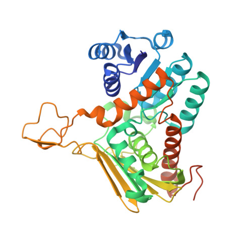
6JW6, 6JW7, 6JW8 - PubMed Abstract:
Kanosamine (3-amino-3-deoxy-d-glucose) is a characteristic sugar unit found in kanamycins, a group of aminoglycoside antibiotics. The kanosamine moiety originates from d-glucose in kanamycin biosynthesis. However, the timing of the replacement of the 3-OH group of the d-glucose-derived biosynthetic intermediate with the amino group is elusive. Comparison of biosynthetic gene clusters for related aminoglycoside antibiotics suggests that the nicotinamide adenine dinucleotide (NAD + )-dependent dehydrogenase KanD2 and the pyridoxal 5'-phosphate (PLP)-dependent aminotransferase KanS2 are responsible for the introduction of the amino group at the C3 position of kanosamine. In this study, we demonstrated that KanD2 and KanS2 convert kanamycin A, B, and C to the corresponding 3″-deamino-3″-hydroxykanamycins (3″-hks) in the presence of PLP, 2-oxoglutarate, and NADH via a reverse reaction in the pathway. Furthermore, we observed that all of the 3″-hks are oxidized by KanD2 with NAD + , but d-glucose, UDP-d-glucose, d-glucose 6-phosphate, and d-glucose 1-phosphate are not. Crystal structure analysis of KanD2 complexed with 3″-hkB and NADH illustrated the selective recognition of pseudotrisaccharides, especially the d-glucose moiety with 2-deoxystreptamine, by a combination of hydrogen bonds and CH-π interactions. Overall, it was clarified that the kanosamine moiety of kanamycins is constructed after the glucosylation of the pseudodisaccharide biosynthetic intermediates in kanamycin biosynthesis.
Organizational Affiliation:
Department of Chemistry, Tokyo Institute of Technology, 2-12-1 O-okayama, Meguro-ku, Tokyo 152-8551, Japan.