6JSB

Crystal structure of Heme binding protein HtaB from Corynebacterium glutamicum


Experimental Data Snapshot

  • Method: X-RAY DIFFRACTION
  • Resolution: 1.70 Å
  • R-Value Free: 0.198 
  • R-Value Work: 0.177 
  • R-Value Observed: 0.178 

wwPDB Validation   3D Report Full Report


Ligand Structure Quality Assessment 


This is version 1.2 of the entry. See complete history


Literature

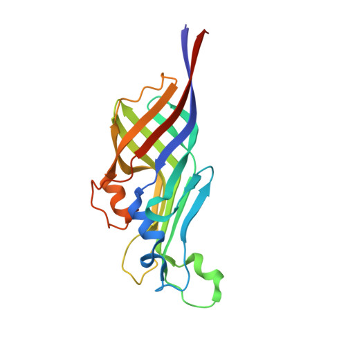
Structural basis for the heme transfer reaction in heme uptake machinery from Corynebacteria.

Muraki, N.Kitatsuji, C.Okamoto, Y.Uchida, T.Ishimori, K.Aono, S.

(2019) Chem Commun (Camb) 55: 13864-13867

  • DOI: https://doi.org/10.1039/c9cc07369h
  • Primary Citation of Related Structures:  
    6JS9, 6JSA, 6JSB, 6JSC, 6JSD

  • PubMed Abstract: 

    The crystal structures of the conserved region domains of HtaA and HtaB, which act as heme binding/transport proteins in the heme uptake machinery in Corynebacterium glutamicum, are determined for the first time. The molecular mechanism of heme transfer among these proteins is proposed based on the spectroscopic and structural analyses.


  • Organizational Affiliation

    Department of Creative Research, Exploratory Research Center on Life and Living Systems (ExCELLS), National Institutes of Natural Sciences, 5-1 Higashiyama, Myodaiji-cho, Okazaki 444-8787, Japan. aono@ims.ac.jp.


Macromolecules
Find similar proteins by:  (by identity cutoff)  |  3D Structure
Entity ID: 1
MoleculeChains Sequence LengthOrganismDetailsImage
Hypothetical membrane protein
A, B
203Corynebacterium glutamicum ATCC 13032Mutation(s): 0 
Gene Names: Cgl0392
UniProt
Find proteins for Q8NTB5 (Corynebacterium glutamicum (strain ATCC 13032 / DSM 20300 / BCRC 11384 / JCM 1318 / LMG 3730 / NCIMB 10025))
Explore Q8NTB5 
Go to UniProtKB:  Q8NTB5
Entity Groups  
Sequence Clusters30% Identity50% Identity70% Identity90% Identity95% Identity100% Identity
UniProt GroupQ8NTB5
Sequence Annotations
Expand
  • Reference Sequence
Experimental Data & Validation

Experimental Data

  • Method: X-RAY DIFFRACTION
  • Resolution: 1.70 Å
  • R-Value Free: 0.198 
  • R-Value Work: 0.177 
  • R-Value Observed: 0.178 
  • Space Group: C 1 2 1
Unit Cell:
Length ( Å )Angle ( ˚ )
a = 235.17α = 90
b = 33.73β = 121.14
c = 126.62γ = 90
Software Package:
Software NamePurpose
PHENIXrefinement
XDSdata reduction
XSCALEdata scaling
PHASERphasing

Structure Validation

View Full Validation Report


Ligand Structure Quality Assessment 


Entry History & Funding Information

Deposition Data


Funding OrganizationLocationGrant Number
Japan Society for the Promotion of ScienceJapan17K15081

Revision History  (Full details and data files)

  • Version 1.0: 2019-11-20
    Type: Initial release
  • Version 1.1: 2019-11-27
    Changes: Database references
  • Version 1.2: 2023-11-22
    Changes: Data collection, Database references, Derived calculations, Refinement description