6JO0

Crystal structure of the DTS-motif rhodopsin from Phaeocystis globosa virus 12T


Experimental Data Snapshot

  • Method: X-RAY DIFFRACTION
  • Resolution: 1.65 Å
  • R-Value Free: 0.209 
  • R-Value Work: 0.192 
  • R-Value Observed: 0.193 

wwPDB Validation   3D Report Full Report


Ligand Structure Quality Assessment 


This is version 1.4 of the entry. See complete history


Literature

A distinct lineage of giant viruses brings a rhodopsin photosystem to unicellular marine predators.

Needham, D.M.Yoshizawa, S.Hosaka, T.Poirier, C.Choi, C.J.Hehenberger, E.Irwin, N.A.T.Wilken, S.Yung, C.M.Bachy, C.Kurihara, R.Nakajima, Y.Kojima, K.Kimura-Someya, T.Leonard, G.Malmstrom, R.R.Mende, D.R.Olson, D.K.Sudo, Y.Sudek, S.Richards, T.A.DeLong, E.F.Keeling, P.J.Santoro, A.E.Shirouzu, M.Iwasaki, W.Worden, A.Z.

(2019) Proc Natl Acad Sci U S A 116: 20574-20583

  • DOI: https://doi.org/10.1073/pnas.1907517116
  • Primary Citation of Related Structures:  
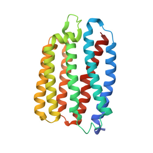
    6JO0

  • PubMed Abstract: 

    Giant viruses are remarkable for their large genomes, often rivaling those of small bacteria, and for having genes thought exclusive to cellular life. Most isolated to date infect nonmarine protists, leaving their strategies and prevalence in marine environments largely unknown. Using eukaryotic single-cell metagenomics in the Pacific, we discovered a Mimiviridae lineage of giant viruses, which infects choanoflagellates, widespread protistan predators related to metazoans. The ChoanoVirus genomes are the largest yet from pelagic ecosystems, with 442 of 862 predicted proteins lacking known homologs. They are enriched in enzymes for modifying organic compounds, including degradation of chitin, an abundant polysaccharide in oceans, and they encode 3 divergent type-1 rhodopsins (VirR) with distinct evolutionary histories from those that capture sunlight in cellular organisms. One (VirR DTS ) is similar to the only other putative rhodopsin from a virus (PgV) with a known host (a marine alga). Unlike the algal virus, ChoanoViruses encode the entire pigment biosynthesis pathway and cleavage enzyme for producing the required chromophore, retinal. We demonstrate that the rhodopsin shared by ChoanoViruses and PgV binds retinal and pumps protons. Moreover, our 1.65-Å resolved VirR DTS crystal structure and mutational analyses exposed differences from previously characterized type-1 rhodopsins, all of which come from cellular organisms. Multiple VirR types are present in metagenomes from across surface oceans, where they are correlated with and nearly as abundant as a canonical marker gene from Mimiviridae Our findings indicate that light-dependent energy transfer systems are likely common components of giant viruses of photosynthetic and phagotrophic unicellular marine eukaryotes.


  • Organizational Affiliation

    Monterey Bay Aquarium Research Institute, Moss Landing, CA 95039.


Macromolecules
Find similar proteins by:  (by identity cutoff)  |  3D Structure
Entity ID: 1
MoleculeChains Sequence LengthOrganismDetailsImage
VirRDTS232Phaeocystis globosa virus 12TMutation(s): 0 
Gene Names: PGAG_00290
Membrane Entity: Yes 
UniProt
Find proteins for G8DI69 (Phaeocystis globosa virus 12T)
Explore G8DI69 
Go to UniProtKB:  G8DI69
Entity Groups  
Sequence Clusters30% Identity50% Identity70% Identity90% Identity95% Identity100% Identity
UniProt GroupG8DI69
Sequence Annotations
Expand
  • Reference Sequence
Small Molecules
Ligands 8 Unique
IDChains Name / Formula / InChI Key2D Diagram3D Interactions
OLB
Query on OLB

Download Ideal Coordinates CCD File 
T [auth A](2S)-2,3-dihydroxypropyl (9Z)-octadec-9-enoate
C21 H40 O4
RZRNAYUHWVFMIP-QJRAZLAKSA-N
RET
Query on RET

Download Ideal Coordinates CCD File 
B [auth A]RETINAL
C20 H28 O
NCYCYZXNIZJOKI-OVSJKPMPSA-N
D12
Query on D12

Download Ideal Coordinates CCD File 
P [auth A]DODECANE
C12 H26
SNRUBQQJIBEYMU-UHFFFAOYSA-N
D10
Query on D10

Download Ideal Coordinates CCD File 
O [auth A]DECANE
C10 H22
DIOQZVSQGTUSAI-UHFFFAOYSA-N
DD9
Query on DD9

Download Ideal Coordinates CCD File 
Q [auth A],
R [auth A],
S [auth A]
nonane
C9 H20
BKIMMITUMNQMOS-UHFFFAOYSA-N
OCT
Query on OCT

Download Ideal Coordinates CCD File 
C [auth A]
D [auth A]
E [auth A]
F [auth A]
G [auth A]
C [auth A],
D [auth A],
E [auth A],
F [auth A],
G [auth A],
H [auth A]
N-OCTANE
C8 H18
TVMXDCGIABBOFY-UHFFFAOYSA-N
PO4
Query on PO4

Download Ideal Coordinates CCD File 
I [auth A]PHOSPHATE ION
O4 P
NBIIXXVUZAFLBC-UHFFFAOYSA-K
HEX
Query on HEX

Download Ideal Coordinates CCD File 
J [auth A],
K [auth A],
L [auth A],
M [auth A],
N [auth A]
HEXANE
C6 H14
VLKZOEOYAKHREP-UHFFFAOYSA-N
Experimental Data & Validation

Experimental Data

  • Method: X-RAY DIFFRACTION
  • Resolution: 1.65 Å
  • R-Value Free: 0.209 
  • R-Value Work: 0.192 
  • R-Value Observed: 0.193 
  • Space Group: P 21 2 21
Unit Cell:
Length ( Å )Angle ( ˚ )
a = 45.194α = 90
b = 57.832β = 90
c = 117.037γ = 90
Software Package:
Software NamePurpose
Aimlessdata scaling
PHENIXrefinement
PDB_EXTRACTdata extraction
XDSdata reduction
PHASERphasing

Structure Validation

View Full Validation Report


Ligand Structure Quality Assessment 


Entry History 

Deposition Data

Revision History  (Full details and data files)

  • Version 1.0: 2019-10-02
    Type: Initial release
  • Version 1.1: 2019-10-09
    Changes: Data collection, Database references
  • Version 1.2: 2019-10-16
    Changes: Data collection, Database references, Structure summary
  • Version 1.3: 2019-10-23
    Changes: Data collection, Database references
  • Version 1.4: 2023-11-22
    Changes: Data collection, Database references, Refinement description