6JC0

Structural analysis of molybdopterin synthases from two mycobacteria pathogens


Experimental Data Snapshot

  • Method: X-RAY DIFFRACTION
  • Resolution: 2.10 Å
  • R-Value Free: 0.237 
  • R-Value Work: 0.190 
  • R-Value Observed: 0.193 

wwPDB Validation   3D Report Full Report


This is version 1.3 of the entry. See complete history


Literature

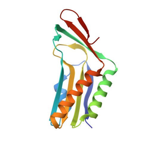
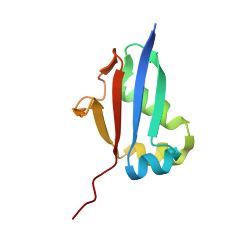
Structural analysis of molybdopterin synthases from two mycobacterial pathogens.

Wang, H.Chen, X.Zhang, W.Zhou, W.Liu, X.Rao, Z.

(2019) Biochem Biophys Res Commun 511: 21-27

  • DOI: https://doi.org/10.1016/j.bbrc.2019.02.024
  • Primary Citation of Related Structures:  
    6JBZ, 6JC0

  • PubMed Abstract: 

    The molybdenum cofactor, composed of molybdopterin and molybdenum, is a necessary compound for the catalytic activity of molybdenum enzymes. Molybdenum cofactor biosynthesis is a conserved multi-step process involving several enzymes. Molybdopterin synthase, a hetero-tetrameric enzyme composed of a pair of MoaE-MoaD subunits, catalyzes the generation of the cis-dithiolene group of molybdopterin in the second step of the process. The cis-dithiolene group can covalently bind molybdenum. Most mycobacterial species possess several genes encoding the full pathway of molybdenum cofactor biosynthesis. In M. smegmatis, the moaD2 and moaE2 genes encode the functional molybdopterin synthase. However, M. tuberculosis has genes encoding several molybdopterin synthase subunit homologs, including moaD1, moaD2, moaE1, moaE2, and moaX, which encodes a MoaD-MoaE fusion protein. Previous studies have shown that moaD2 and moaE2 encode functional molybdopterin synthase. Here, we report the crystal structures of two substrate-free molybdopterin synthases from two different mycobacterial pathogens, M. tuberculosis and M. smegmatis, at 2.1 Å and 2.6 Å resolutions, respectively. The overall structure of both molybdopterin synthases was hetero-tetrameric, consisting of a MoaE2 dimer flanked on either side by single MoaD2 subunits. The carboxyl-terminal domain of MoaD2 inserted into MoaE2, forming the active pocket. A comparison with previously reported molybdopterin synthase structures showed that substrate-binding and catalytic residues were conserved, despite low sequence similarity among these enzymes. The low sequence identity at the MoaE-MoaD heterodimer interface may provide the structural basis to explore mycobacterial inhibitors.


  • Organizational Affiliation

    State Key Laboratory of Medicinal Chemical Biology and College of Pharmacy, Nankai University, Tianjin, China; College of Life Science, Nankai University, Tianjin, China.


Macromolecules
Find similar proteins by:  (by identity cutoff)  |  3D Structure
Entity ID: 1
MoleculeChains Sequence LengthOrganismDetailsImage
Putative molybdenum cofactor biosynthesis proteinA [auth B],
C [auth D]
142Mycolicibacterium smegmatis MC2 155Mutation(s): 0 
Gene Names: moaE2MSMEI_5550
UniProt
Find proteins for I7FL16 (Mycolicibacterium smegmatis (strain ATCC 700084 / mc(2)155))
Explore I7FL16 
Go to UniProtKB:  I7FL16
Entity Groups  
Sequence Clusters30% Identity50% Identity70% Identity90% Identity95% Identity100% Identity
UniProt GroupI7FL16
Sequence Annotations
Expand
  • Reference Sequence
Find similar proteins by:  (by identity cutoff)  |  3D Structure
Entity ID: 2
MoleculeChains Sequence LengthOrganismDetailsImage
Putative molybdenum cofactor biosynthesis protein D2 (MoaD2) / thiamine SB [auth C],
D [auth A]
88Mycolicibacterium smegmatis MC2 155Mutation(s): 0 
Gene Names: moaD2MSMEI_5548
UniProt
Find proteins for I7FT00 (Mycolicibacterium smegmatis (strain ATCC 700084 / mc(2)155))
Explore I7FT00 
Go to UniProtKB:  I7FT00
Entity Groups  
Sequence Clusters30% Identity50% Identity70% Identity90% Identity95% Identity100% Identity
UniProt GroupI7FT00
Sequence Annotations
Expand
  • Reference Sequence
Experimental Data & Validation

Experimental Data

  • Method: X-RAY DIFFRACTION
  • Resolution: 2.10 Å
  • R-Value Free: 0.237 
  • R-Value Work: 0.190 
  • R-Value Observed: 0.193 
  • Space Group: P 1 21 1
Unit Cell:
Length ( Å )Angle ( ˚ )
a = 62.763α = 90
b = 57.599β = 99.29
c = 66.577γ = 90
Software Package:
Software NamePurpose
PHENIXrefinement
HKL-2000data reduction
HKL-2000data scaling
PHASERphasing

Structure Validation

View Full Validation Report


Entry History & Funding Information

Deposition Data


Funding OrganizationLocationGrant Number
National Natural Science Foundation of ChinaChinaGrant Nos. 813300237 and 81520108019

Revision History  (Full details and data files)

  • Version 1.0: 2019-02-13
    Type: Initial release
  • Version 1.1: 2019-03-06
    Changes: Data collection, Database references
  • Version 1.2: 2019-03-13
    Changes: Data collection, Database references
  • Version 1.3: 2023-11-22
    Changes: Data collection, Database references, Refinement description