6J9R

Coiled-coil Domain of Drosophila TRIM Protein Brat


Experimental Data Snapshot

  • Method: X-RAY DIFFRACTION
  • Resolution: 2.50 Å
  • R-Value Free: 0.269 
  • R-Value Work: 0.224 
  • R-Value Observed: 0.226 

wwPDB Validation   3D Report Full Report


This is version 1.1 of the entry. See complete history


Literature

Crystal structure of the coiled-coil domain of Drosophila TRIM protein Brat.

Liu, C.Shan, Z.Diao, J.Wen, W.Wang, W.

(2019) Proteins 87: 706-710

  • DOI: https://doi.org/10.1002/prot.25691
  • Primary Citation of Related Structures:  
    6J9R

  • PubMed Abstract: 

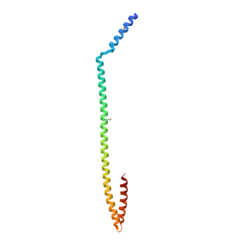
    Drosophila brain tumor (Brat) is a translational repressor belonging to the tripartite motif (TRIM) protein superfamily. During the asymmetric division of Drosophila neuroblasts, Brat localizes at the basal cortex via direct interaction with the scaffolding protein Miranda (Mira), and segregates into the basal ganglion mother cells after cell division. It was previously reported that both the coiled-coil (CC) and NHL domains of Brat are required for the interaction with Mira, but the underlying structural basis is elusive. Here, we determine the crystal structure of Brat-CC domain (aa 376-511) at 2.5 Å, showing that Brat-CC forms an elongated antiparallel dimer through an unconventional CC structure. The dimeric assembly in Brat-CC structure is similar to its counterparts in other TRIM proteins, but Brat-CC also exhibits some distinct structural features. We also demonstrate that the CC domain could not bind Mira by its own, neither does the isolated NHL domain of Brat. Rather, Brat binds to Mira through the CC-NHL domain tandem, indicating that the function of the CC domain is to assemble Brat-NHL in dimeric form, which is necessary for Mira binding.


  • Organizational Affiliation

    Department of Chemistry, Institutes of Biomedical Sciences and Multiscale Research Institute of Complex System, Fudan University, Shanghai, People's Republic of China.


Macromolecules
Find similar proteins by:  (by identity cutoff)  |  3D Structure
Entity ID: 1
MoleculeChains Sequence LengthOrganismDetailsImage
Brain tumor protein
A, B
136Drosophila melanogaster American nodavirus (ANV) SW-2009aMutation(s): 0 
UniProt
Find proteins for Q8MQJ9 (Drosophila melanogaster)
Explore Q8MQJ9 
Go to UniProtKB:  Q8MQJ9
Entity Groups  
Sequence Clusters30% Identity50% Identity70% Identity90% Identity95% Identity100% Identity
UniProt GroupQ8MQJ9
Sequence Annotations
Expand
  • Reference Sequence
Small Molecules
Ligands 1 Unique
IDChains Name / Formula / InChI Key2D Diagram3D Interactions
GOL
Query on GOL

Download Ideal Coordinates CCD File 
C [auth A]GLYCEROL
C3 H8 O3
PEDCQBHIVMGVHV-UHFFFAOYSA-N
Modified Residues  1 Unique
IDChains TypeFormula2D DiagramParent
MSE
Query on MSE
A, B
L-PEPTIDE LINKINGC5 H11 N O2 SeMET
Experimental Data & Validation

Experimental Data

  • Method: X-RAY DIFFRACTION
  • Resolution: 2.50 Å
  • R-Value Free: 0.269 
  • R-Value Work: 0.224 
  • R-Value Observed: 0.226 
  • Space Group: P 21 21 21
Unit Cell:
Length ( Å )Angle ( ˚ )
a = 51.612α = 90
b = 61.1β = 90
c = 128.084γ = 90
Software Package:
Software NamePurpose
HKL-2000data reduction
HKL-2000data scaling
PHASERphasing
PHENIXrefinement
PDB_EXTRACTdata extraction

Structure Validation

View Full Validation Report


Entry History 

Deposition Data

Revision History  (Full details and data files)

  • Version 1.0: 2019-04-24
    Type: Initial release
  • Version 1.1: 2019-07-17
    Changes: Data collection, Database references