6J4N

Structure of papua new guinea MBL-1(PNGM-1) native


Experimental Data Snapshot

  • Method: X-RAY DIFFRACTION
  • Resolution: 2.10 Å
  • R-Value Free: 0.244 
  • R-Value Work: 0.190 
  • R-Value Observed: 0.193 

wwPDB Validation   3D Report Full Report


This is version 1.2 of the entry. See complete history


Literature

The novel metallo-beta-lactamase PNGM-1 from a deep-sea sediment metagenome: crystallization and X-ray crystallographic analysis.

Park, K.S.Hong, M.K.Jeon, J.W.Kim, J.H.Jeon, J.H.Lee, J.H.Kim, T.Y.Karim, A.M.Malik, S.K.Kang, L.W.Lee, S.H.

(2018) Acta Crystallogr F Struct Biol Commun 74: 644-649

  • DOI: https://doi.org/10.1107/S2053230X18012268
  • Primary Citation of Related Structures:  
    6J4N

  • PubMed Abstract: 

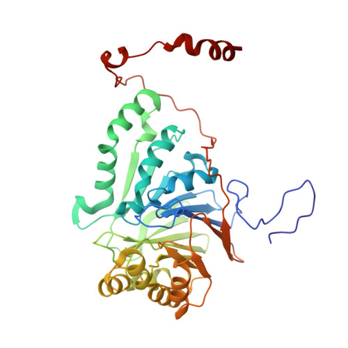
    Metallo-β-lactamases (MBLs) are present in major Gram-negative pathogens and environmental species, and pose great health risks because of their ability to hydrolyze the β-lactam rings of antibiotics such as carbapenems. PNGM-1 was the first reported case of a subclass B3 MBL protein that was identified from a metagenomic library from deep-sea sediments that predate the antibiotic era. In this study, PNGM-1 was overexpressed, purified and crystallized. Crystals of native and selenomethionine-substituted PNGM-1 diffracted to 2.10 and 2.30 Å resolution, respectively. Both the native and the selenomethionine-labelled PNGM-1 crystals belonged to the monoclinic space group P2 1 , with unit-cell parameters a = 122, b = 83, c = 163 Å, β = 110°. Matthews coefficient (V M ) calculations suggested the presence of 6-10 molecules in the asymmetric unit, corresponding to a solvent content of ∼31-58%. Structure determination is currently in progress.


  • Organizational Affiliation

    National Leading Research Laboratory of Drug Resistance Proteomics, Department of Biological Sciences, Myongji University, 116 Myongjiro, Yongin, Gyeonggido 17058, Republic of Korea.


Macromolecules
Find similar proteins by:  (by identity cutoff)  |  3D Structure
Entity ID: 1
MoleculeChains Sequence LengthOrganismDetailsImage
Metallo-beta-lactamases PNGM-1
A, B, C, D, E
A, B, C, D, E, F, G, H
372uncultured bacteriumMutation(s): 0 
Entity Groups  
Sequence Clusters30% Identity50% Identity70% Identity90% Identity95% Identity100% Identity
Sequence Annotations
Expand
  • Reference Sequence
Small Molecules
Ligands 1 Unique
IDChains Name / Formula / InChI Key2D Diagram3D Interactions
ZN
Query on ZN

Download Ideal Coordinates CCD File 
I [auth A]
J [auth A]
K [auth B]
L [auth B]
M [auth C]
I [auth A],
J [auth A],
K [auth B],
L [auth B],
M [auth C],
N [auth C],
O [auth D],
P [auth D],
Q [auth E],
R [auth E],
S [auth F],
T [auth F],
U [auth G],
V [auth G],
W [auth H],
X [auth H]
ZINC ION
Zn
PTFCDOFLOPIGGS-UHFFFAOYSA-N
Experimental Data & Validation

Experimental Data

  • Method: X-RAY DIFFRACTION
  • Resolution: 2.10 Å
  • R-Value Free: 0.244 
  • R-Value Work: 0.190 
  • R-Value Observed: 0.193 
  • Space Group: P 1 21 1
Unit Cell:
Length ( Å )Angle ( ˚ )
a = 122.274α = 90
b = 83.017β = 110.55
c = 163.469γ = 90
Software Package:
Software NamePurpose
REFMACrefinement
HKL-2000data collection
HKL-2000data reduction
MOLREPphasing

Structure Validation

View Full Validation Report


Entry History 

Deposition Data

Revision History  (Full details and data files)

  • Version 1.0: 2019-02-27
    Type: Initial release
  • Version 1.1: 2019-05-22
    Changes: Data collection, Source and taxonomy
  • Version 1.2: 2024-03-27
    Changes: Data collection, Database references, Derived calculations