6IYY

Crystal structure of human WIPI3,loop deletion mutant


Experimental Data Snapshot

  • Method: X-RAY DIFFRACTION
  • Resolution: 1.80 Å
  • R-Value Free: 0.204 
  • R-Value Work: 0.165 
  • R-Value Observed: 0.168 

wwPDB Validation   3D Report Full Report


This is version 1.2 of the entry. See complete history


Literature

Structural Conservation of the Two Phosphoinositide-Binding Sites in WIPI Proteins.

Liang, R.Ren, J.Zhang, Y.Feng, W.

(2019) J Mol Biol 431: 1494-1505

  • DOI: https://doi.org/10.1016/j.jmb.2019.02.019
  • Primary Citation of Related Structures:  
    6IYY

  • PubMed Abstract: 

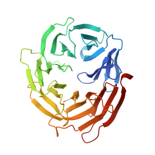
    WIPI proteins are mammalian PROPPIN family members that bind to phosphoinositides and play prominent roles in autophagosome biogenesis. Two phosphoinositide-binding sites were previously described in yeast PROPPIN Hsv2 but remain to be determined in mammalian WIPI proteins. Here, we characterized four human WIPI proteins (WIPI1-4) and solved the structure of WIPI3. WIPI proteins can bind to PI(3)P and PI(3,5)P 2 and adopt a conventional seven-bladed β-propeller fold. The structure of WIPI3 revealed that WIPI proteins also contain two sites embedded in blades 5 and 6 for recognizing phosphoinositides, resembling that in Hsv2. Structural comparison further demonstrated that the two conserved phosphoinositide-binding sites in PROPPIN proteins are not identical but intrinsically tend to recognize different types of phosphoinositides. This work provides the structural evidence to support the conservation of the two phosphoinositide-binding sites in WIPI proteins and also uncovers the potential phosphoinositide-binding selectivity for each site.


  • Organizational Affiliation

    National Laboratory of Biomacromolecules, CAS Center for Excellence in Biomacromolecules, Institute of Biophysics, Chinese Academy of Sciences, 15 Datun Road, Beijing 100101, China; College of Life Sciences, University of Chinese Academy of Sciences, Beijing 100049, China.


Macromolecules
Find similar proteins by:  (by identity cutoff)  |  3D Structure
Entity ID: 1
MoleculeChains Sequence LengthOrganismDetailsImage
WD repeat domain phosphoinositide-interacting protein 3317Homo sapiensMutation(s): 0 
Gene Names: WIPI3
UniProt & NIH Common Fund Data Resources
Find proteins for Q5MNZ6 (Homo sapiens)
Explore Q5MNZ6 
Go to UniProtKB:  Q5MNZ6
PHAROS:  Q5MNZ6
GTEx:  ENSG00000141580 
Entity Groups  
Sequence Clusters30% Identity50% Identity70% Identity90% Identity95% Identity100% Identity
UniProt GroupQ5MNZ6
Sequence Annotations
Expand
  • Reference Sequence
Experimental Data & Validation

Experimental Data

  • Method: X-RAY DIFFRACTION
  • Resolution: 1.80 Å
  • R-Value Free: 0.204 
  • R-Value Work: 0.165 
  • R-Value Observed: 0.168 
  • Space Group: C 2 2 21
Unit Cell:
Length ( Å )Angle ( ˚ )
a = 46.297α = 90
b = 104.931β = 90
c = 124.882γ = 90
Software Package:
Software NamePurpose
HKL-2000data reduction
HKL-2000data scaling
PDB_EXTRACTdata extraction
PHENIXrefinement
AutoSolphasing

Structure Validation

View Full Validation Report


Entry History & Funding Information

Deposition Data


Funding OrganizationLocationGrant Number
National Natural Science Foundation of ChinaChina31470746
National Natural Science Foundation of ChinaChina31770786
National Natural Science Foundation of ChinaChina31600608
National Natural Science Foundation of ChinaChina31600622

Revision History  (Full details and data files)

  • Version 1.0: 2019-03-13
    Type: Initial release
  • Version 1.1: 2019-04-10
    Changes: Data collection, Database references
  • Version 1.2: 2024-03-27
    Changes: Data collection, Database references