6IXD

X-ray crystal structure of bPI-11 hiv-1 protease complex


Experimental Data Snapshot

  • Method: X-RAY DIFFRACTION
  • Resolution: 1.00 Å
  • R-Value Free: 0.150 
  • R-Value Work: 0.132 
  • R-Value Observed: 0.133 

wwPDB Validation   3D Report Full Report


Ligand Structure Quality Assessment 


This is version 1.2 of the entry. See complete history


Literature

Acquired Removability of Aspartic Protease Inhibitors by Direct Biotinylation.

Hidaka, K.Adachi, M.Tsuda, Y.

(2019) Bioconjug Chem 30: 1979-1985

  • DOI: https://doi.org/10.1021/acs.bioconjchem.9b00195
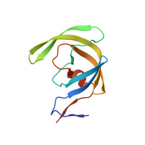
  • Primary Citation of Related Structures:  
    6IXD

  • PubMed Abstract: 

    Protease inhibitors are used as both research tools and therapeutics. Many of these inhibitors consist of substrate amino acid sequence-derived structure with a transition state mimic to interact with the active site of the protease, suppressing enzymatic activity. However, once they bind, macrodilution or protein denaturation is required to remove them, limiting their usage. In this study, we describe a removable protease inhibitor, which is a directly biotinylated analogue to control the activities of HIV-1 protease and human cathepsin D. In the substrate cleavage assay, we observed that the nanomolar inhibitory activities were lost upon the addition of streptavidin, while the enzymatic activities sufficiently recovered. HIV-1 protease mixed with the removable inhibitor, avoiding autolysis, was still active to be detected by adding streptavidin after one year at room temperature. We also observed that the inhibitor was an effective eluent for the simple detection of the activity of proteases purified from human serum and cells. These results demonstrate that direct biotinylation of protease inhibitors could be a novel method for controlling the enzymatic activity from OFF to ON. We proposed the phenomenon that binding equilibrium of inhibitor was shifted from protease to streptavidin with higher affinity, named "inhibitor stripping action by affinity competition", or ISAAC. We anticipate that ISAAC could be applicable for preservatives of proteases and activity-based diagnosis of protease related diseases. Furthermore, removable inhibitor to be designed for targeted proteases changing the inhibitor structure may elucidate enzymatic activity in intrinsic form with natural modifications from various biological samples.


  • Organizational Affiliation

    Institute for Quantum Life Science , National Institutes for Quantum and Radiological Science and Technology , Tokai, Ibaraki , 319-1106 , Japan.


Macromolecules
Find similar proteins by:  (by identity cutoff)  |  3D Structure
Entity ID: 1
MoleculeChains Sequence LengthOrganismDetailsImage
Protease
A, B
99Human immunodeficiency virus 1Mutation(s): 5 
Gene Names: pol
EC: 3.4.23.16
UniProt
Find proteins for Q5RZ08 (Human immunodeficiency virus 1)
Explore Q5RZ08 
Go to UniProtKB:  Q5RZ08
Entity Groups  
Sequence Clusters30% Identity50% Identity70% Identity90% Identity95% Identity100% Identity
UniProt GroupQ5RZ08
Sequence Annotations
Expand
  • Reference Sequence
Small Molecules
Ligands 4 Unique
IDChains Name / Formula / InChI Key2D Diagram3D Interactions
B0F
Query on B0F

Download Ideal Coordinates CCD File 
E [auth B](4R)-3-[(2S,3S)-3-[2-[4-[5-[(3aS,4S,6aR)-2-oxidanylidene-1,3,3a,4,6,6a-hexahydrothieno[3,4-d]imidazol-4-yl]pentanoylamino]-2,6-dimethyl-phenoxy]ethanoylamino]-2-oxidanyl-4-phenyl-butanoyl]-5,5-dimethyl-N-[(1S,2R)-2-oxidanyl-2,3-dihydro-1H-inden-1-yl]-1,3-thiazolidine-4-carboxamide
C45 H56 N6 O8 S2
FGRIDPZSJUPACI-SOOZJBRBSA-N
SO4
Query on SO4

Download Ideal Coordinates CCD File 
D [auth A],
H [auth B]
SULFATE ION
O4 S
QAOWNCQODCNURD-UHFFFAOYSA-L
GOL
Query on GOL

Download Ideal Coordinates CCD File 
C [auth A],
F [auth B],
G [auth B]
GLYCEROL
C3 H8 O3
PEDCQBHIVMGVHV-UHFFFAOYSA-N
CL
Query on CL

Download Ideal Coordinates CCD File 
I [auth B]CHLORIDE ION
Cl
VEXZGXHMUGYJMC-UHFFFAOYSA-M
Binding Affinity Annotations 
IDSourceBinding Affinity
B0F Binding MOAD:  6IXD Ki: 0.16 (nM) from 1 assay(s)
Experimental Data & Validation

Experimental Data

  • Method: X-RAY DIFFRACTION
  • Resolution: 1.00 Å
  • R-Value Free: 0.150 
  • R-Value Work: 0.132 
  • R-Value Observed: 0.133 
  • Space Group: P 21 21 2
Unit Cell:
Length ( Å )Angle ( ˚ )
a = 58.302α = 90
b = 86.01β = 90
c = 46.549γ = 90
Software Package:
Software NamePurpose
PHENIXrefinement
HKL-2000data reduction
SCALEPACKdata scaling
PHENIXphasing

Structure Validation

View Full Validation Report


Ligand Structure Quality Assessment 


Entry History 

Deposition Data

Revision History  (Full details and data files)

  • Version 1.0: 2019-05-22
    Type: Initial release
  • Version 1.1: 2019-07-31
    Changes: Data collection, Database references
  • Version 1.2: 2023-11-22
    Changes: Data collection, Database references, Refinement description