6IUJ

Crystal structure of GH30 xylanase B from Talaromyces cellulolyticus


Experimental Data Snapshot

  • Method: X-RAY DIFFRACTION
  • Resolution: 2.25 Å
  • R-Value Free: 0.202 
  • R-Value Work: 0.166 
  • R-Value Observed: 0.168 

wwPDB Validation   3D Report Full Report


Ligand Structure Quality Assessment 


This is version 2.1 of the entry. See complete history


Literature

Structural and functional characterization of a bifunctional GH30-7 xylanase B from the filamentous fungusTalaromyces cellulolyticus.

Nakamichi, Y.Fouquet, T.Ito, S.Watanabe, M.Matsushika, A.Inoue, H.

(2019) J Biol Chem 294: 4065-4078

  • DOI: https://doi.org/10.1074/jbc.RA118.007207
  • Primary Citation of Related Structures:  
    6IUJ

  • PubMed Abstract: 

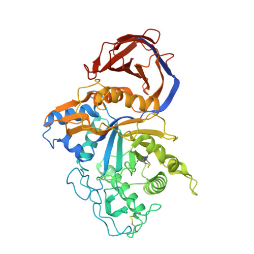
    Glucuronoxylanases are endo-xylanases and members of the glycoside hydrolase family 30 subfamilies 7 (GH30-7) and 8 (GH30-8). Unlike for the well-studied GH30-8 enzymes, the structural and functional characteristics of GH30-7 enzymes remain poorly understood. Here, we report the catalytic properties and three-dimensional structure of GH30-7 xylanase B (Xyn30B) identified from the cellulolytic fungus Talaromyces cellulolyticus Xyn30B efficiently degraded glucuronoxylan to acidic xylooligosaccharides (XOSs), including an α-1,2-linked 4- O -methyl-d-glucuronosyl substituent (MeGlcA). Rapid analysis with negative-mode electrospray-ionization multistage MS (ESI(-)-MS n ) revealed that the structures of the acidic XOS products are the same as those of the hydrolysates (MeGlcA 2 Xyl n , n > 2) obtained with typical glucuronoxylanases. Acidic XOS products were further degraded by Xyn30B, releasing first xylobiose and then xylotetraose and xylohexaose as transglycosylation products. This hydrolase reaction was unique to Xyn30B, and the substrate was cleaved at the xylobiose unit from its nonreducing end, indicating that Xyn30B is a bifunctional enzyme possessing both endo-glucuronoxylanase and exo-xylobiohydrolase activities. The crystal structure of Xyn30B was determined as the first structure of a GH30-7 xylanase at 2.25 Å resolution, revealing that Xyn30B is composed of a pseudo-(α/β) 8 -catalytic domain, lacking an α6 helix, and a small β-rich domain. This structure and site-directed mutagenesis clarified that Arg 46 , conserved in GH30-7 glucuronoxylanases, is a critical residue for MeGlcA appendage-dependent xylan degradation. The structural comparison between Xyn30B and the GH30-8 enzymes suggests that Asn 93 in the β2-α2 loop is involved in xylobiohydrolase activity. In summary, our findings indicate that Xyn30B is a bifunctional endo- and exo-xylanase.


  • Organizational Affiliation

    From the Bioconversion Group and.


Macromolecules
Find similar proteins by:  (by identity cutoff)  |  3D Structure
Entity ID: 1
MoleculeChains Sequence LengthOrganismDetailsImage
GH30 Xylanase B
A, B
474Talaromyces pinophilus CF-2612Mutation(s): 0 
UniProt
Find proteins for A0A4V8H018 (Talaromyces pinophilus CF-2612)
Explore A0A4V8H018 
Go to UniProtKB:  A0A4V8H018
Entity Groups  
Sequence Clusters30% Identity50% Identity70% Identity90% Identity95% Identity100% Identity
UniProt GroupA0A4V8H018
Sequence Annotations
Expand
  • Reference Sequence
Oligosaccharides

Help

Entity ID: 2
MoleculeChains Length2D Diagram Glycosylation3D Interactions
2-acetamido-2-deoxy-beta-D-glucopyranose-(1-4)-2-acetamido-2-deoxy-beta-D-glucopyranose
C, H, K
2N-Glycosylation
Glycosylation Resources
GlyTouCan:  G42666HT
GlyCosmos:  G42666HT
GlyGen:  G42666HT
Entity ID: 3
MoleculeChains Length2D Diagram Glycosylation3D Interactions
alpha-D-mannopyranose-(1-3)-[alpha-D-mannopyranose-(1-6)]beta-D-mannopyranose-(1-4)-2-acetamido-2-deoxy-beta-D-glucopyranose-(1-4)-2-acetamido-2-deoxy-beta-D-glucopyranose
D
5N-Glycosylation
Glycosylation Resources
GlyTouCan:  G22768VO
GlyCosmos:  G22768VO
GlyGen:  G22768VO
Entity ID: 4
MoleculeChains Length2D Diagram Glycosylation3D Interactions
beta-D-mannopyranose-(1-4)-2-acetamido-2-deoxy-beta-D-glucopyranose-(1-4)-2-acetamido-2-deoxy-beta-D-glucopyranose
E, F, I
3N-Glycosylation
Glycosylation Resources
GlyTouCan:  G15407YE
GlyCosmos:  G15407YE
GlyGen:  G15407YE
Entity ID: 5
MoleculeChains Length2D Diagram Glycosylation3D Interactions
alpha-D-mannopyranose-(1-6)-beta-D-mannopyranose-(1-4)-2-acetamido-2-deoxy-beta-D-glucopyranose-(1-4)-2-acetamido-2-deoxy-beta-D-glucopyranose
G
4N-Glycosylation
Glycosylation Resources
GlyTouCan:  G22573RC
GlyCosmos:  G22573RC
GlyGen:  G22573RC
Entity ID: 6
MoleculeChains Length2D Diagram Glycosylation3D Interactions
alpha-D-mannopyranose-(1-3)-alpha-D-mannopyranose-(1-6)-[alpha-D-mannopyranose-(1-3)]beta-D-mannopyranose-(1-4)-2-acetamido-2-deoxy-beta-D-glucopyranose-(1-4)-2-acetamido-2-deoxy-beta-D-glucopyranose
J
6N-Glycosylation
Glycosylation Resources
GlyTouCan:  G09724ZC
GlyCosmos:  G09724ZC
GlyGen:  G09724ZC
Experimental Data & Validation

Experimental Data

  • Method: X-RAY DIFFRACTION
  • Resolution: 2.25 Å
  • R-Value Free: 0.202 
  • R-Value Work: 0.166 
  • R-Value Observed: 0.168 
  • Space Group: P 21 21 21
Unit Cell:
Length ( Å )Angle ( ˚ )
a = 83.172α = 90
b = 114.948β = 90
c = 118.461γ = 90
Software Package:
Software NamePurpose
REFMACrefinement
XDSdata reduction
XDSdata scaling
MOLREPphasing

Structure Validation

View Full Validation Report


Ligand Structure Quality Assessment 


Entry History 

Deposition Data

Revision History  (Full details and data files)

  • Version 1.0: 2019-01-30
    Type: Initial release
  • Version 1.1: 2019-03-27
    Changes: Data collection, Database references
  • Version 2.0: 2020-07-29
    Type: Remediation
    Reason: Carbohydrate remediation
    Changes: Atomic model, Data collection, Derived calculations, Structure summary
  • Version 2.1: 2023-11-22
    Changes: Data collection, Database references, Derived calculations, Refinement description, Structure summary