6ILZ

Crystal structure of PKCiota in complex with inhibitor


Experimental Data Snapshot

  • Method: X-RAY DIFFRACTION
  • Resolution: 3.26 Å
  • R-Value Free: 0.348 
  • R-Value Work: 0.306 
  • R-Value Observed: 0.310 

wwPDB Validation   3D Report Full Report


Ligand Structure Quality Assessment 


This is version 1.1 of the entry. See complete history


Literature

Fragment-based Discovery of a Small-Molecule Protein Kinase C-iota Inhibitor Binding Post-kinase Domain Residues.

Kwiatkowski, J.Baburajendran, N.Poulsen, A.Liu, B.Tee, D.H.Y.Wong, Y.X.Poh, Z.Y.Ong, E.H.Dinie, N.Cherian, J.Jansson, A.E.Hill, J.Keller, T.H.Hung, A.W.

(2019) ACS Med Chem Lett 10: 318-323

  • DOI: https://doi.org/10.1021/acsmedchemlett.8b00546
  • Primary Citation of Related Structures:  
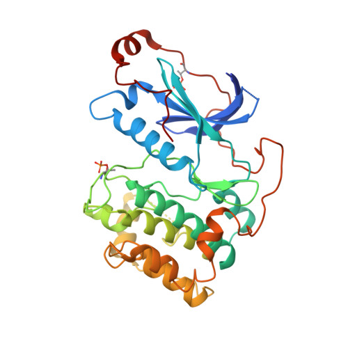
    6ILZ

  • PubMed Abstract: 

    The atypical protein kinase C-iota (PKC-ι) enzyme is implicated in various cancers and has been put forward as an attractive target for developing anticancer therapy. A high concentration biochemical screen identified pyridine fragment weakly inhibiting PKC-ι with IC 50 = 424 μM. Driven by structure-activity relationships and guided by docking hypothesis, the weakly bound fragment was eventually optimized into a potent inhibitor of PKC-ι (IC 50 = 270 nM). Through the course of the optimization, an intermediate compound was crystallized with the protein, and careful analysis of the X-ray crystal structure revealed a unique binding mode involving the post-kinase domain (C-terminal tail) of PKC-ι.


  • Organizational Affiliation

    Experimental Therapeutics Centre, Agency for Science, Technology and Research (ASTAR), 11 Biopolis Way, Helios #03-10/11, Singapore 138667, Singapore.


Macromolecules
Find similar proteins by:  (by identity cutoff)  |  3D Structure
Entity ID: 1
MoleculeChains Sequence LengthOrganismDetailsImage
Protein kinase C iota typeA,
B [auth C],
C [auth E],
D [auth G]
345Homo sapiensMutation(s): 0 
Gene Names: PRKCIDXS1179E
EC: 2.7.11.13
UniProt & NIH Common Fund Data Resources
Find proteins for P41743 (Homo sapiens)
Explore P41743 
Go to UniProtKB:  P41743
PHAROS:  P41743
GTEx:  ENSG00000163558 
Entity Groups  
Sequence Clusters30% Identity50% Identity70% Identity90% Identity95% Identity100% Identity
UniProt GroupP41743
Sequence Annotations
Expand
  • Reference Sequence
Small Molecules
Modified Residues  1 Unique
IDChains TypeFormula2D DiagramParent
TPO
Query on TPO
A,
B [auth C],
C [auth E],
D [auth G]
L-PEPTIDE LINKINGC4 H10 N O6 PTHR
Experimental Data & Validation

Experimental Data

  • Method: X-RAY DIFFRACTION
  • Resolution: 3.26 Å
  • R-Value Free: 0.348 
  • R-Value Work: 0.306 
  • R-Value Observed: 0.310 
  • Space Group: P 1 21 1
Unit Cell:
Length ( Å )Angle ( ˚ )
a = 87.87α = 90
b = 87.63β = 114.69
c = 105.181γ = 90
Software Package:
Software NamePurpose
PHENIXrefinement
PDB_EXTRACTdata extraction
Cootmodel building
XDSdata scaling
PHENIXphasing

Structure Validation

View Full Validation Report


Ligand Structure Quality Assessment 


Entry History 

Deposition Data

Revision History  (Full details and data files)

  • Version 1.0: 2019-06-26
    Type: Initial release
  • Version 1.1: 2023-11-22
    Changes: Data collection, Database references, Refinement description