


Crystal structure of fructuronate-tagaturonate epimerase UxaE from Cohnella laeviribosi
Choi, M.Y., Kang, L.W., Ho, T.H., Nguyen, D.Q., Lee, I.H., Lee, J.H., Park, Y.S., Park, H.J.To be published.
Experimental Data Snapshot
wwPDB Validation 3D Report Full Report

Entity ID: 1 | |||||
|---|---|---|---|---|---|
| Molecule | Chains | Sequence Length | Organism | Details | Image |
| Fructuronate-tagaturonate epimerase UxaE | A, B [auth D] | 489 | Cohnella laeviribosi | Mutation(s): 0 EC: 5.1.2.7 | 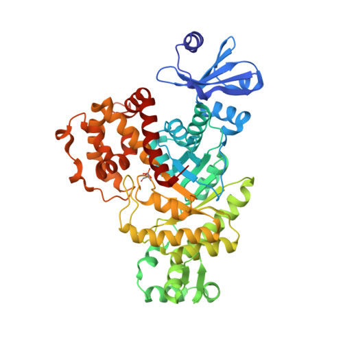 |
Entity Groups | |||||
| Sequence Clusters | 30% Identity50% Identity70% Identity90% Identity95% Identity100% Identity | ||||
Sequence AnnotationsExpand | |||||
| |||||
| Ligands 2 Unique | |||||
|---|---|---|---|---|---|
| ID | Chains | Name / Formula / InChI Key | 2D Diagram | 3D Interactions | |
| EDO Query on EDO | D [auth A], F [auth D] | 1,2-ETHANEDIOL C2 H6 O2 LYCAIKOWRPUZTN-UHFFFAOYSA-N |  | ||
| MN Query on MN | C [auth A], E [auth D] | MANGANESE (II) ION Mn WAEMQWOKJMHJLA-UHFFFAOYSA-N |  | ||
| Modified Residues 1 Unique | |||||
|---|---|---|---|---|---|
| ID | Chains | Type | Formula | 2D Diagram | Parent |
| SEP Query on SEP | A, B [auth D] | L-PEPTIDE LINKING | C3 H8 N O6 P |  | SER |
| Length ( Å ) | Angle ( ˚ ) |
|---|---|
| a = 51.816 | α = 98.14 |
| b = 73.47 | β = 110.16 |
| c = 74.673 | γ = 90.12 |
| Software Name | Purpose |
|---|---|
| REFMAC | refinement |
| HKL-2000 | data scaling |
| HKL-2000 | data collection |
| HKL-2000 | data reduction |
| MOLREP | phasing |
RCSB PDB (citation) is hosted by
RCSB PDB is a member of the

