


Structural Insights into Phosphite Dehydrogenase Variants Favoring a Non-natural Redox Cofactor
Liu, Y., Feng, Y., Wang, L., Guo, X., Liu, W., Li, Q., Wang, X., Xue, S., Zhao, Z.(2019) ACS Catal 9: 1883-1887
Experimental Data Snapshot
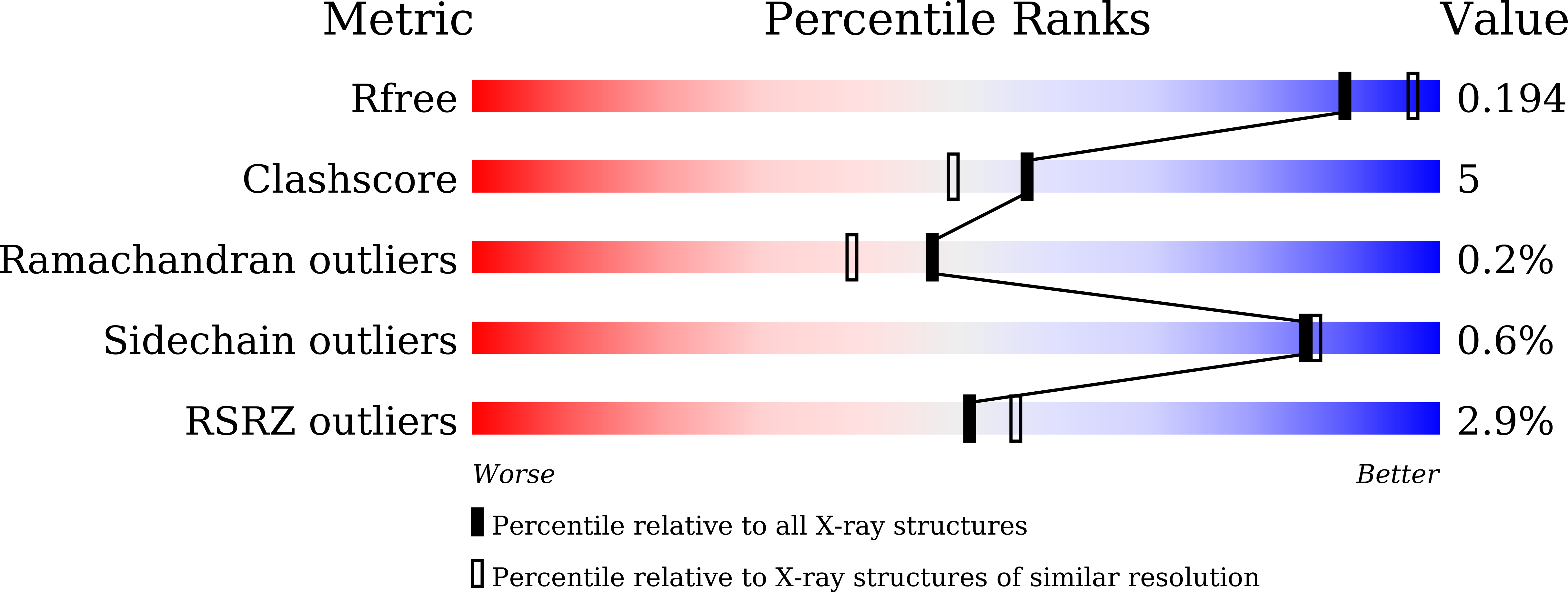
wwPDB Validation 3D Report Full Report
Entity ID: 1 | |||||
|---|---|---|---|---|---|
| Molecule | Chains | Sequence Length | Organism | Details | Image |
| Phosphite dehydrogenase | 336 | Ralstonia sp. 4506 | Mutation(s): 0 Gene Names: ptxD | 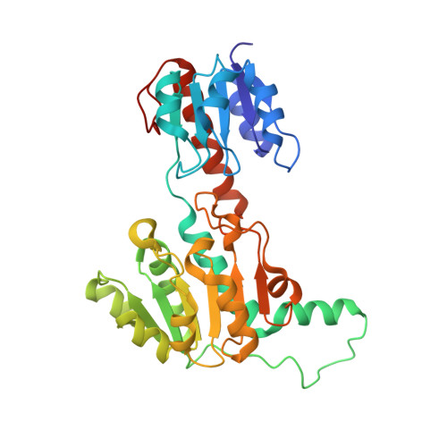 | |
UniProt | |||||
Find proteins for G4XDR8 (Ralstonia sp. 4506) Explore G4XDR8 Go to UniProtKB: G4XDR8 | |||||
Entity Groups | |||||
| Sequence Clusters | 30% Identity50% Identity70% Identity90% Identity95% Identity100% Identity | ||||
| UniProt Group | G4XDR8 | ||||
Sequence AnnotationsExpand | |||||
| |||||
| Length ( Å ) | Angle ( ˚ ) |
|---|---|
| a = 61.653 | α = 90 |
| b = 97.849 | β = 90 |
| c = 122.44 | γ = 90 |
| Software Name | Purpose |
|---|---|
| PHENIX | refinement |
| HKL-2000 | data reduction |
| HKL-2000 | data scaling |
| PHENIX | phasing |
RCSB PDB (citation) is hosted by
RCSB PDB is a member of the

