6IFD

Crystal Structure of CMP-N-acetylneuraminate Synthetase from Vibrio cholerae in complex with CDP and Mg2+.


Experimental Data Snapshot

  • Method: X-RAY DIFFRACTION
  • Resolution: 2.30 Å
  • R-Value Free: 0.240 
  • R-Value Work: 0.202 
  • R-Value Observed: 0.203 

wwPDB Validation   3D Report Full Report


Ligand Structure Quality Assessment 


This is version 1.1 of the entry. See complete history


Literature

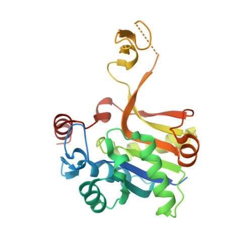
Structural and functional characterization of CMP-N-acetylneuraminate synthetase from Vibrio cholerae.

Bose, S.Purkait, D.Joseph, D.Nayak, V.Subramanian, R.

(2019) Acta Crystallogr D Struct Biol 75: 564-577

  • DOI: https://doi.org/10.1107/S2059798319006831
  • Primary Citation of Related Structures:  
    6IFD, 6IFI

  • PubMed Abstract: 

    Several pathogenic bacteria utilize sialic acid, including host-derived N-acetylneuraminic acid (Neu5Ac), in at least two ways: they use it as a nutrient source and as a host-evasion strategy by coating themselves with Neu5Ac. Given the significant role of sialic acid in pathogenesis and host-gut colonization by various pathogenic bacteria, including Neisseria meningitidis, Haemophilus influenzae, Pasteurella multocida and Vibrio cholerae, several enzymes of the sialic acid catabolic, biosynthetic and incorporation pathways are considered to be potential drug targets. In this work, findings on the structural and functional characterization of CMP-N-acetylneuraminate synthetase (CMAS), a key enzyme in the incorporation pathway, from Vibrio cholerae are reported. CMAS catalyzes the synthesis of CMP-sialic acid by utilizing CTP and sialic acid. Crystal structures of the apo and the CDP-bound forms of the enzyme were determined, which allowed the identification of the metal cofactor Mg 2+ in the active site interacting with CDP and the invariant Asp215 residue. While open and closed structural forms of the enzyme from eukaryotic and other bacterial species have already been characterized, a partially closed structure of V. cholerae CMAS (VcCMAS) observed upon CDP binding, representing an intermediate state, is reported here. The kinetic data suggest that VcCMAS is capable of activating the two most common sialic acid derivatives, Neu5Ac and Neu5Gc. Amino-acid sequence and structural comparison of the active site of VcCMAS with those of eukaryotic and other bacterial counterparts reveal a diverse hydrophobic pocket that interacts with the C5 substituents of sialic acid. Analyses of the thermodynamic signatures obtained from the binding of the nucleotide (CTP) and the product (CMP-sialic acid) to VcCMAS provide fundamental information on the energetics of the binding process.


  • Organizational Affiliation

    Institute for Stem Cell Science and Regenerative Medicine, GKVK Post, Bellary Road, Bangalore 560 065, India.


Macromolecules
Find similar proteins by:  (by identity cutoff)  |  3D Structure
Entity ID: 1
MoleculeChains Sequence LengthOrganismDetailsImage
CMP-N-acetylneuraminate Synthetase
A, B, C, D
251Vibrio choleraeMutation(s): 0 
EC: 2.7.7.43
Entity Groups  
Sequence Clusters30% Identity50% Identity70% Identity90% Identity95% Identity100% Identity
Sequence Annotations
Expand
  • Reference Sequence
Small Molecules
Ligands 6 Unique
IDChains Name / Formula / InChI Key2D Diagram3D Interactions
CDP
Query on CDP

Download Ideal Coordinates CCD File 
E [auth A],
H [auth B],
K [auth C]
CYTIDINE-5'-DIPHOSPHATE
C9 H15 N3 O11 P2
ZWIADYZPOWUWEW-XVFCMESISA-N
PG4
Query on PG4

Download Ideal Coordinates CCD File 
M [auth C]TETRAETHYLENE GLYCOL
C8 H18 O5
UWHCKJMYHZGTIT-UHFFFAOYSA-N
PGE
Query on PGE

Download Ideal Coordinates CCD File 
F [auth A]TRIETHYLENE GLYCOL
C6 H14 O4
ZIBGPFATKBEMQZ-UHFFFAOYSA-N
EDO
Query on EDO

Download Ideal Coordinates CCD File 
G [auth A],
J [auth B],
N [auth D]
1,2-ETHANEDIOL
C2 H6 O2
LYCAIKOWRPUZTN-UHFFFAOYSA-N
CA
Query on CA

Download Ideal Coordinates CCD File 
I [auth B]CALCIUM ION
Ca
BHPQYMZQTOCNFJ-UHFFFAOYSA-N
MG
Query on MG

Download Ideal Coordinates CCD File 
L [auth C]MAGNESIUM ION
Mg
JLVVSXFLKOJNIY-UHFFFAOYSA-N
Experimental Data & Validation

Experimental Data

  • Method: X-RAY DIFFRACTION
  • Resolution: 2.30 Å
  • R-Value Free: 0.240 
  • R-Value Work: 0.202 
  • R-Value Observed: 0.203 
  • Space Group: P 21 21 21
Unit Cell:
Length ( Å )Angle ( ˚ )
a = 93.38α = 90
b = 97.72β = 90
c = 98.76γ = 90
Software Package:
Software NamePurpose
BUSTERrefinement
PDB_EXTRACTdata extraction
XDSdata reduction
XSCALEdata scaling
PHASERphasing

Structure Validation

View Full Validation Report


Ligand Structure Quality Assessment 


Entry History & Funding Information

Deposition Data


Funding OrganizationLocationGrant Number
IndiaBT/IN/SWEDEN/06/SR/2017-2018
IndiaBT/PR5081/INF/156/2012
IndiaBT/PR12422/MED/31/287/214

Revision History  (Full details and data files)

  • Version 1.0: 2019-07-03
    Type: Initial release
  • Version 1.1: 2023-11-22
    Changes: Data collection, Database references, Derived calculations, Refinement description