6I2P

Crystal structure of the Mycobacterium tuberculosis PknB kinase domain (L33E mutant) in complex with its substrate GarA


Experimental Data Snapshot

  • Method: X-RAY DIFFRACTION
  • Resolution: 2.37 Å
  • R-Value Free: 0.234 
  • R-Value Work: 0.220 
  • R-Value Observed: 0.221 

wwPDB Validation   3D Report Full Report


Ligand Structure Quality Assessment 


This is version 1.1 of the entry. See complete history


Literature

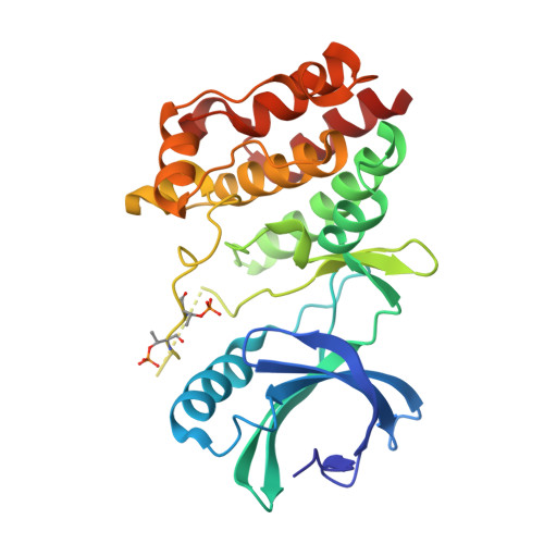
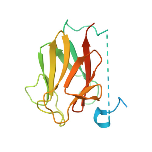
Structural insights into the functional versatility of an FHA domain protein in mycobacterial signaling.

Wagner, T.Andre-Leroux, G.Hindie, V.Barilone, N.Lisa, M.N.Hoos, S.Raynal, B.Vulliez-Le Normand, B.O'Hare, H.M.Bellinzoni, M.Alzari, P.M.

(2019) Sci Signal 12

  • DOI: https://doi.org/10.1126/scisignal.aav9504
  • Primary Citation of Related Structures:  
    6I2P, 6I2Q, 6I2R, 6I2S

  • PubMed Abstract: 

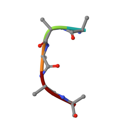
    Forkhead-associated (FHA) domains are modules that bind to phosphothreonine (pThr) residues in signaling cascades. The FHA-containing mycobacterial protein GarA is a central element of a phosphorylation-dependent signaling pathway that redirects metabolic flux in response to amino acid starvation or cell growth requirements. GarA acts as a phosphorylation-dependent ON/OFF molecular switch. In its nonphosphorylated ON state, the GarA FHA domain engages in phosphorylation-independent interactions with various metabolic enzymes that orchestrate nitrogen flow, such as 2-oxoglutarate decarboxylase (KGD). However, phosphorylation at the GarA N-terminal region by the protein kinase PknB or PknG triggers autoinhibition through the intramolecular association of the N-terminal domain with the FHA domain, thus blocking all downstream interactions. To investigate these different FHA binding modes, we solved the crystal structures of the mycobacterial upstream (phosphorylation-dependent) complex PknB-GarA and the downstream (phosphorylation-independent) complex GarA-KGD. Our results show that the phosphorylated activation loop of PknB serves as a docking site to recruit GarA through canonical FHA-pThr interactions. However, the same GarA FHA-binding pocket targets an allosteric site on nonphosphorylated KGD, where a key element of recognition is a phosphomimetic aspartate. Further enzymatic and mutagenesis studies revealed that GarA acted as a dynamic allosteric inhibitor of KGD by preventing crucial motions in KGD that are necessary for catalysis. Our results provide evidence for physiological phosphomimetics, supporting numerous mutagenesis studies using such approaches, and illustrate how evolution can shape a single FHA-binding pocket to specifically interact with multiple phosphorylated and nonphosphorylated protein partners.


  • Organizational Affiliation

    Institut Pasteur, Unité de Microbiologie Structurale, CNRS UMR 3528 & Université Paris Diderot, 25 Rue du Docteur Roux, 75724 Paris Cedex 15, France.


Macromolecules
Find similar proteins by:  (by identity cutoff)  |  3D Structure
Entity ID: 1
MoleculeChains Sequence LengthOrganismDetailsImage
Serine/threonine-protein kinase PknB
A, B
280Mycobacterium tuberculosis H37RvMutation(s): 1 
Gene Names: pknBRv0014cMTCY10H4.14c
EC: 2.7.11.1
UniProt
Find proteins for P9WI81 (Mycobacterium tuberculosis (strain ATCC 25618 / H37Rv))
Explore P9WI81 
Go to UniProtKB:  P9WI81
Entity Groups  
Sequence Clusters30% Identity50% Identity70% Identity90% Identity95% Identity100% Identity
UniProt GroupP9WI81
Sequence Annotations
Expand
  • Reference Sequence
Find similar proteins by:  (by identity cutoff)  |  3D Structure
Entity ID: 2
MoleculeChains Sequence LengthOrganismDetailsImage
Glycogen accumulation regulator GarAC [auth D],
D [auth E]
163Mycobacterium tuberculosis H37RvMutation(s): 0 
Gene Names: garARv1827MTCY1A11.16c
UniProt
Find proteins for P9WJA9 (Mycobacterium tuberculosis (strain ATCC 25618 / H37Rv))
Explore P9WJA9 
Go to UniProtKB:  P9WJA9
Entity Groups  
Sequence Clusters30% Identity50% Identity70% Identity90% Identity95% Identity100% Identity
UniProt GroupP9WJA9
Sequence Annotations
Expand
  • Reference Sequence

Find similar proteins by:  Sequence   |   3D Structure  

Entity ID: 3
MoleculeChains Sequence LengthOrganismDetailsImage
UNK-UNK-UNK-UNK-UNKE [auth C]5Mycobacterium tuberculosis H37RvMutation(s): 0 
Sequence Annotations
Expand
  • Reference Sequence
Small Molecules
Modified Residues  1 Unique
IDChains TypeFormula2D DiagramParent
TPO
Query on TPO
A, B
L-PEPTIDE LINKINGC4 H10 N O6 PTHR
Experimental Data & Validation

Experimental Data

  • Method: X-RAY DIFFRACTION
  • Resolution: 2.37 Å
  • R-Value Free: 0.234 
  • R-Value Work: 0.220 
  • R-Value Observed: 0.221 
  • Space Group: P 21 21 21
Unit Cell:
Length ( Å )Angle ( ˚ )
a = 69.31α = 90
b = 70.46β = 90
c = 188.6γ = 90
Software Package:
Software NamePurpose
BUSTERrefinement
XDSdata reduction
Aimlessdata scaling
PHASERphasing

Structure Validation

View Full Validation Report


Ligand Structure Quality Assessment 


Entry History 

Deposition Data

Revision History  (Full details and data files)

  • Version 1.0: 2019-05-22
    Type: Initial release
  • Version 1.1: 2024-01-24
    Changes: Data collection, Database references, Derived calculations, Refinement description