6I20

Flavin Analogue Sheds Light on Light-Oxygen-Voltage Domain Mechanism


Experimental Data Snapshot

  • Method: X-RAY DIFFRACTION
  • Resolution: 1.37 Å
  • R-Value Free: 0.175 
  • R-Value Work: 0.151 
  • R-Value Observed: 0.152 

wwPDB Validation   3D Report Full Report


Ligand Structure Quality Assessment 


This is version 2.0 of the entry. See complete history


Literature

A Noncanonical Chromophore Reveals Structural Rearrangements of the Light-Oxygen-Voltage Domain upon Photoactivation.

Kalvaitis, M.E.Johnson, L.A.Mart, R.J.Rizkallah, P.Allemann, R.K.

(2019) Biochemistry 58: 2608-2616

  • DOI: https://doi.org/10.1021/acs.biochem.9b00255
  • Primary Citation of Related Structures:  
    6I20, 6I21, 6I22, 6I23, 6I24, 6I25

  • PubMed Abstract: 

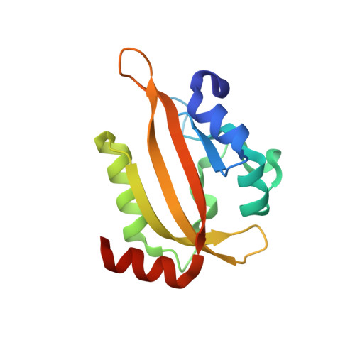
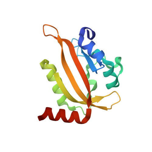
    Light-oxygen-voltage (LOV) domains are increasingly used to engineer photoresponsive biological systems. While the photochemical cycle is well documented, the allosteric mechanism by which formation of a cysteinyl-flavin adduct leads to activation is unclear. Via replacement of flavin mononucleotide (FMN) with 5-deazaflavin mononucleotide (5dFMN) in the Aureochrome1a (Au1a) transcription factor from Ochromonas danica, a thermally stable cysteinyl-5dFMN adduct was generated. High-resolution crystal structures (<2 Å) under different illumination conditions with either FMN or 5dFMN chromophores reveal three conformations of the highly conserved glutamine 293. An allosteric hydrogen bond network linking the chromophore via Gln293 to the auxiliary A'α helix is observed. With FMN, a "flip" of the Gln293 side chain occurs between dark and lit states. 5dFMN cannot hydrogen bond through the C5 position and proved to be unable to support Au1a domain dimerization. Under blue light, the Gln293 side chain instead "swings" away in a conformation distal to the chromophore and not previously observed in existing LOV domain structures. Together, the multiple side chain conformations of Gln293 and functional analysis of 5dFMN provide new insight into the structural requirements for LOV domain activation.


  • Organizational Affiliation

    School of Chemistry , Cardiff University , Park Place , Cardiff CF10 3AT , United Kingdom.


Macromolecules
Find similar proteins by:  (by identity cutoff)  |  3D Structure
Entity ID: 1
MoleculeChains Sequence LengthOrganismDetailsImage
Aureochrome1-like protein
A, C, D
134Ochromonas danicaMutation(s): 0 
Gene Names: OdAUREO1
UniProt
Find proteins for C5NSW6 (Ochromonas danica)
Explore C5NSW6 
Go to UniProtKB:  C5NSW6
Entity Groups  
Sequence Clusters30% Identity50% Identity70% Identity90% Identity95% Identity100% Identity
UniProt GroupC5NSW6
Sequence Annotations
Expand
  • Reference Sequence
Find similar proteins by:  (by identity cutoff)  |  3D Structure
Entity ID: 2
MoleculeChains Sequence LengthOrganismDetailsImage
Aureochrome1-like protein134Ochromonas danicaMutation(s): 0 
Gene Names: OdAUREO1
UniProt
Find proteins for C5NSW6 (Ochromonas danica)
Explore C5NSW6 
Go to UniProtKB:  C5NSW6
Entity Groups  
Sequence Clusters30% Identity50% Identity70% Identity90% Identity95% Identity100% Identity
UniProt GroupC5NSW6
Sequence Annotations
Expand
  • Reference Sequence
Small Molecules
Ligands 5 Unique
IDChains Name / Formula / InChI Key2D Diagram3D Interactions
FMN
Query on FMN

Download Ideal Coordinates CCD File 
E [auth A],
K [auth B],
O [auth C],
Q [auth D]
FLAVIN MONONUCLEOTIDE
C17 H21 N4 O9 P
FVTCRASFADXXNN-SCRDCRAPSA-N
MES
Query on MES

Download Ideal Coordinates CCD File 
M [auth B]2-(N-MORPHOLINO)-ETHANESULFONIC ACID
C6 H13 N O4 S
SXGZJKUKBWWHRA-UHFFFAOYSA-N
EDO
Query on EDO

Download Ideal Coordinates CCD File 
F [auth A]
G [auth A]
H [auth A]
L [auth B]
R [auth D]
F [auth A],
G [auth A],
H [auth A],
L [auth B],
R [auth D],
S [auth D]
1,2-ETHANEDIOL
C2 H6 O2
LYCAIKOWRPUZTN-UHFFFAOYSA-N
ACT
Query on ACT

Download Ideal Coordinates CCD File 
I [auth A]ACETATE ION
C2 H3 O2
QTBSBXVTEAMEQO-UHFFFAOYSA-M
CL
Query on CL

Download Ideal Coordinates CCD File 
J [auth A],
N [auth B],
P [auth C],
T [auth D]
CHLORIDE ION
Cl
VEXZGXHMUGYJMC-UHFFFAOYSA-M
Experimental Data & Validation

Experimental Data

  • Method: X-RAY DIFFRACTION
  • Resolution: 1.37 Å
  • R-Value Free: 0.175 
  • R-Value Work: 0.151 
  • R-Value Observed: 0.152 
  • Space Group: P 21 21 21
Unit Cell:
Length ( Å )Angle ( ˚ )
a = 65.68α = 90
b = 104.04β = 90
c = 105.94γ = 90
Software Package:
Software NamePurpose
REFMACrefinement
XDSdata reduction
Aimlessdata scaling
MOLREPphasing
xia2data reduction

Structure Validation

View Full Validation Report


Ligand Structure Quality Assessment 


Entry History 

Deposition Data

Revision History  (Full details and data files)

  • Version 1.0: 2019-05-29
    Type: Initial release
  • Version 1.1: 2019-06-19
    Changes: Data collection, Database references
  • Version 2.0: 2019-10-02
    Changes: Advisory, Atomic model, Data collection, Database references, Derived calculations, Other, Polymer sequence, Source and taxonomy, Structure summary