6HY7

Crystal structure of alpha9 nAChR extracellular domain in complex with alpha-conotoxin RgIA


Experimental Data Snapshot

  • Method: X-RAY DIFFRACTION
  • Resolution: 2.26 Å
  • R-Value Free: 0.249 
  • R-Value Work: 0.193 
  • R-Value Observed: 0.196 

wwPDB Validation   3D Report Full Report


Ligand Structure Quality Assessment 


This is version 1.4 of the entry. See complete history


Literature

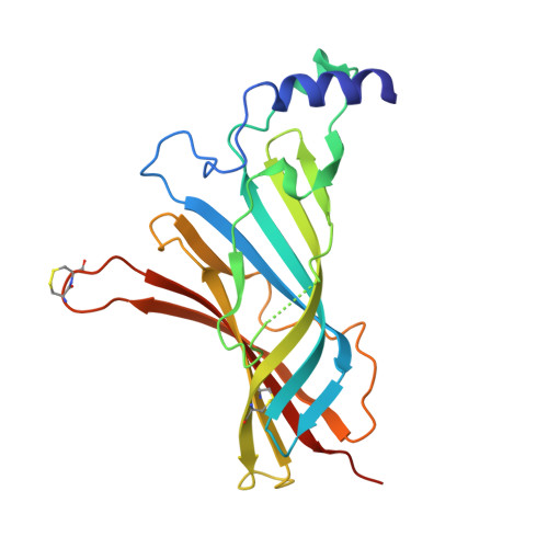
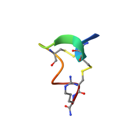
Crystal Structure of the Monomeric Extracellular Domain of alpha 9 Nicotinic Receptor Subunit in Complex With alpha-Conotoxin RgIA: Molecular Dynamics Insights Into RgIA Binding to alpha 9 alpha 10 Nicotinic Receptors.

Zouridakis, M.Papakyriakou, A.Ivanov, I.A.Kasheverov, I.E.Tsetlin, V.Tzartos, S.Giastas, P.

(2019) Front Pharmacol 10: 474-474

  • DOI: https://doi.org/10.3389/fphar.2019.00474
  • Primary Citation of Related Structures:  
    6HY7

  • PubMed Abstract: 

    The α9 subunit of nicotinic acetylcholine receptors (nAChRs) exists mainly in heteropentameric assemblies with α10. Accumulating data indicate the presence of three different binding sites in α9α10 nAChRs: the α9(+)/α9(-), the α9(+)/α10(-), and the α10(+)/α9(-). The major role of the principal (+) side of the extracellular domain (ECD) of α9 subunit in binding of the antagonists methyllylcaconitine and α-bungarotoxin was shown previously by the crystal structures of the monomeric α9-ECD with these molecules. Here we present the 2.26-Å resolution crystal structure of α9-ECD in complex with α-conotoxin (α-Ctx) RgIA, a potential drug for chronic pain, the first structure reported for a complex between an nAChR domain and an α-Ctx. Superposition of this structure with those of other α-Ctxs bound to the homologous pentameric acetylcholine binding proteins revealed significant similarities in the orientation of bound conotoxins, despite the monomeric state of the α9-ECD. In addition, ligand-binding studies calculated a binding affinity of RgIA to the α9-ECD at the low micromolar range. Given the high identity between α9 and α10 ECDs, particularly at their (+) sides, the presented structure was used as template for molecular dynamics simulations of the ECDs of the human α9α10 nAChR in pentameric assemblies. Our results support a favorable binding of RgIA at α9(+)/α9(-) or α10(+)/α9(-) rather than the α9(+)/α10(-) interface, in accordance with previous mutational and functional data.


  • Organizational Affiliation

    Department of Neurobiology, Hellenic Pasteur Institute, Athens, Greece.


Macromolecules
Find similar proteins by:  (by identity cutoff)  |  3D Structure
Entity ID: 1
MoleculeChains Sequence LengthOrganismDetailsImage
Neuronal acetylcholine receptor subunit alpha-9218Homo sapiensMutation(s): 0 
Gene Names: CHRNA9NACHRA9
UniProt & NIH Common Fund Data Resources
Find proteins for Q9UGM1 (Homo sapiens)
Explore Q9UGM1 
Go to UniProtKB:  Q9UGM1
PHAROS:  Q9UGM1
GTEx:  ENSG00000174343 
Entity Groups  
Sequence Clusters30% Identity50% Identity70% Identity90% Identity95% Identity100% Identity
UniProt GroupQ9UGM1
Sequence Annotations
Expand
  • Reference Sequence

Find similar proteins by:  Sequence   |   3D Structure  

Entity ID: 2
MoleculeChains Sequence LengthOrganismDetailsImage
Alpha-conotoxin RgIA13Conus regiusMutation(s): 0 
UniProt
Find proteins for P0C1D0 (Conus regius)
Explore P0C1D0 
Go to UniProtKB:  P0C1D0
Entity Groups  
Sequence Clusters30% Identity50% Identity70% Identity90% Identity95% Identity100% Identity
UniProt GroupP0C1D0
Sequence Annotations
Expand
  • Reference Sequence
Small Molecules
Modified Residues  1 Unique
IDChains TypeFormula2D DiagramParent
AAR
Query on AAR
B
L-PEPTIDE LINKINGC6 H16 N5 OARG
Experimental Data & Validation

Experimental Data

  • Method: X-RAY DIFFRACTION
  • Resolution: 2.26 Å
  • R-Value Free: 0.249 
  • R-Value Work: 0.193 
  • R-Value Observed: 0.196 
  • Space Group: P 21 21 2
Unit Cell:
Length ( Å )Angle ( ˚ )
a = 63.646α = 90
b = 82.53β = 90
c = 49.474γ = 90
Software Package:
Software NamePurpose
PHENIXrefinement
XDSdata reduction
XDSdata scaling
PHASERphasing

Structure Validation

View Full Validation Report


Ligand Structure Quality Assessment 


Entry History 

Deposition Data

Revision History  (Full details and data files)

  • Version 1.0: 2019-05-22
    Type: Initial release
  • Version 1.1: 2019-06-05
    Changes: Data collection, Database references
  • Version 1.2: 2019-08-21
    Changes: Data collection
  • Version 1.3: 2020-07-29
    Type: Remediation
    Reason: Carbohydrate remediation
    Changes: Data collection, Derived calculations, Structure summary
  • Version 1.4: 2024-01-24
    Changes: Data collection, Database references, Refinement description, Structure summary