6HUY

HmdII from Desulfurobacterium thermolithotrophum reconstitued with Fe-guanylylpyridinol (FeGP) cofactor and co-crystallized with methenyl-tetrahydrofolate form A


Experimental Data Snapshot

  • Method: X-RAY DIFFRACTION
  • Resolution: 2.25 Å
  • R-Value Free: 0.212 
  • R-Value Work: 0.187 
  • R-Value Observed: 0.188 

wwPDB Validation   3D Report Full Report


Ligand Structure Quality Assessment 


This is version 1.3 of the entry. See complete history


Literature

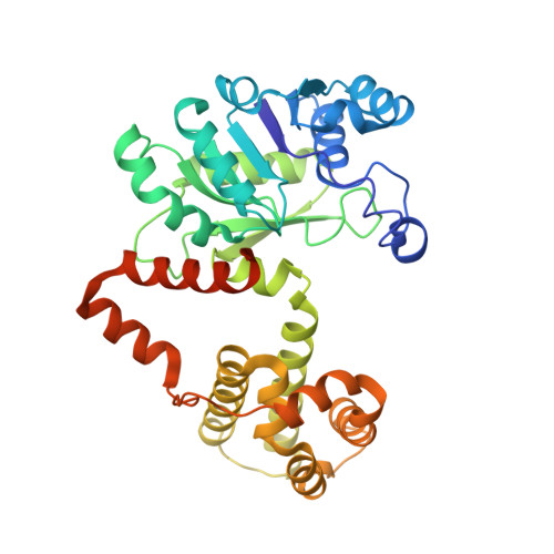
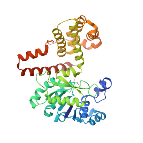
The Bacterial [Fe]-Hydrogenase Paralog HmdII Uses Tetrahydrofolate Derivatives as Substrates.

Watanabe, T.Wagner, T.Huang, G.Kahnt, J.Ataka, K.Ermler, U.Shima, S.

(2019) Angew Chem Int Ed Engl 58: 3506-3510

  • DOI: https://doi.org/10.1002/anie.201813465
  • Primary Citation of Related Structures:  
    6HUX, 6HUY, 6HUZ

  • PubMed Abstract: 

    [Fe]-hydrogenase (Hmd) catalyzes the reversible hydrogenation of methenyl-tetrahydromethanopterin (methenyl-H 4 MPT + ) with H 2 . H 4 MPT is a C1-carrier of methanogenic archaea. One bacterial genus, Desulfurobacterium, contains putative genes for the Hmd paralog, termed HmdII, and the HcgA-G proteins. The latter are required for the biosynthesis of the prosthetic group of Hmd, the iron-guanylylpyridinol (FeGP) cofactor. This finding is intriguing because Hmd and HmdII strictly use H 4 MPT derivatives that are absent in most bacteria. We identified the presence of the FeGP cofactor in D. thermolithotrophum. The bacterial HmdII reconstituted with the FeGP cofactor catalyzed the hydrogenation of derivatives of tetrahydrofolate, the bacterial C1-carrier, albeit with low enzymatic activities. The crystal structures show how Hmd recognizes tetrahydrofolate derivatives. These findings have an impact on future biotechnology by identifying a bacterial Hmd paralog.


  • Organizational Affiliation

    Microbial Protein Structure Group, Max Planck Institute for Terrestrial Microbiology, Karl-von-Frisch Straße 10, 35043, Marburg, Germany.


Macromolecules
Find similar proteins by:  (by identity cutoff)  |  3D Structure
Entity ID: 1
MoleculeChains Sequence LengthOrganismDetailsImage
Coenzyme F420-dependent N(5),N(10)-methenyltetrahydromethanopterin reductase-related protein
A, C
370Desulfurobacterium thermolithotrophum DSM 11699Mutation(s): 0 
Gene Names: Dester_1504
EC: 1.12.98.2
UniProt
Find proteins for F0S2B6 (Desulfurobacterium thermolithotrophum (strain DSM 11699 / BSA))
Explore F0S2B6 
Go to UniProtKB:  F0S2B6
Entity Groups  
Sequence Clusters30% Identity50% Identity70% Identity90% Identity95% Identity100% Identity
UniProt GroupF0S2B6
Sequence Annotations
Expand
  • Reference Sequence
Find similar proteins by:  (by identity cutoff)  |  3D Structure
Entity ID: 2
MoleculeChains Sequence LengthOrganismDetailsImage
Coenzyme F420-dependent N(5),N(10)-methenyltetrahydromethanopterin reductase-related protein
B, D
370Desulfurobacterium thermolithotrophum DSM 11699Mutation(s): 0 
Gene Names: Dester_1504
UniProt
Find proteins for F0S2B6 (Desulfurobacterium thermolithotrophum (strain DSM 11699 / BSA))
Explore F0S2B6 
Go to UniProtKB:  F0S2B6
Entity Groups  
Sequence Clusters30% Identity50% Identity70% Identity90% Identity95% Identity100% Identity
UniProt GroupF0S2B6
Sequence Annotations
Expand
  • Reference Sequence
Small Molecules
Ligands 5 Unique
IDChains Name / Formula / InChI Key2D Diagram3D Interactions
FE9
Query on FE9

Download Ideal Coordinates CCD File 
E [auth A],
N [auth C]
iron-guanylyl pyridinol cofactor
C21 H23 Fe N6 O13 P S
AEHOAZNVUAGELD-VPXBKTNXSA-K
GUE
Query on GUE

Download Ideal Coordinates CCD File 
G [auth A],
M [auth B],
Q [auth C],
V [auth D]
5,10-Methenyltetrahydrofolate
C20 H22 N7 O6
MEANFMOQMXYMCT-OLZOCXBDSA-O
PG4
Query on PG4

Download Ideal Coordinates CCD File 
L [auth B]TETRAETHYLENE GLYCOL
C8 H18 O5
UWHCKJMYHZGTIT-UHFFFAOYSA-N
GOL
Query on GOL

Download Ideal Coordinates CCD File 
F [auth A],
R [auth D]
GLYCEROL
C3 H8 O3
PEDCQBHIVMGVHV-UHFFFAOYSA-N
DMS
Query on DMS

Download Ideal Coordinates CCD File 
H [auth B]
I [auth B]
J [auth B]
K [auth B]
O [auth C]
H [auth B],
I [auth B],
J [auth B],
K [auth B],
O [auth C],
P [auth C],
S [auth D],
T [auth D],
U [auth D]
DIMETHYL SULFOXIDE
C2 H6 O S
IAZDPXIOMUYVGZ-UHFFFAOYSA-N
Modified Residues  1 Unique
IDChains TypeFormula2D DiagramParent
CSO
Query on CSO
B, D
L-PEPTIDE LINKINGC3 H7 N O3 SCYS
Experimental Data & Validation

Experimental Data

  • Method: X-RAY DIFFRACTION
  • Resolution: 2.25 Å
  • R-Value Free: 0.212 
  • R-Value Work: 0.187 
  • R-Value Observed: 0.188 
  • Space Group: P 21 21 21
Unit Cell:
Length ( Å )Angle ( ˚ )
a = 99.769α = 90
b = 131.829β = 90
c = 168.263γ = 90
Software Package:
Software NamePurpose
PHENIXrefinement
XDSdata reduction
SCALAdata scaling
PHASERphasing

Structure Validation

View Full Validation Report


Ligand Structure Quality Assessment 


Entry History & Funding Information

Deposition Data


Funding OrganizationLocationGrant Number
Max Planck SocietyGermany--
Ministry of Education (China)China--

Revision History  (Full details and data files)

  • Version 1.0: 2019-01-09
    Type: Initial release
  • Version 1.1: 2019-02-06
    Changes: Data collection, Database references
  • Version 1.2: 2019-03-13
    Changes: Data collection, Database references
  • Version 1.3: 2024-01-24
    Changes: Data collection, Database references, Derived calculations, Refinement description