6HUL

Sulfolobus solfataricus Tryptophan Synthase AB Complex


Experimental Data Snapshot

  • Method: X-RAY DIFFRACTION
  • Resolution: 2.55 Å
  • R-Value Free: 0.239 
  • R-Value Work: 0.182 
  • R-Value Observed: 0.185 

wwPDB Validation   3D Report Full Report


Ligand Structure Quality Assessment 


This is version 2.3 of the entry. See complete history


Literature

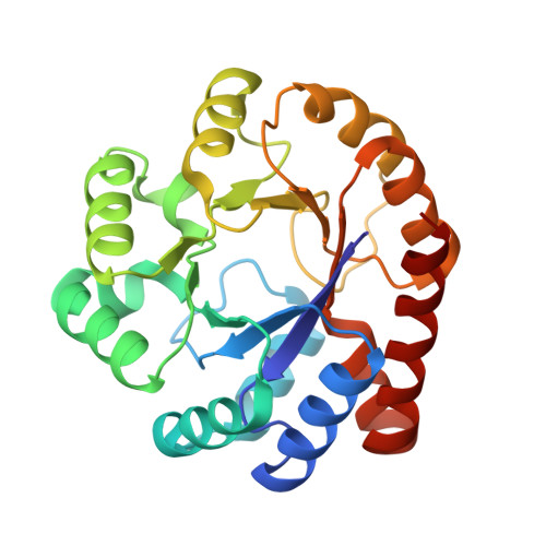
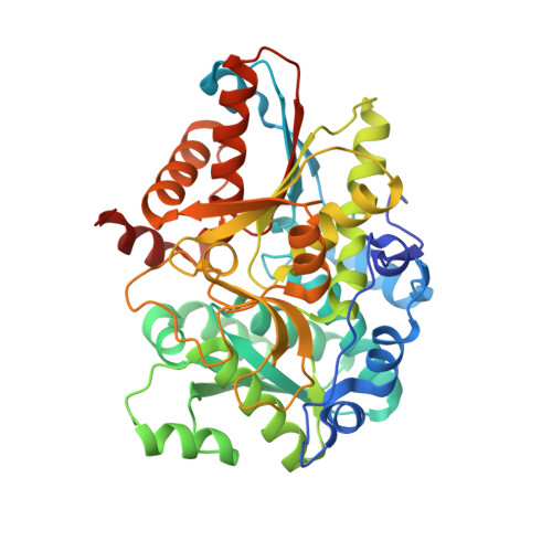
Evolutionary Morphing of Tryptophan Synthase: Functional Mechanisms for the Enzymatic Channeling of Indole.

Fleming, J.R.Schupfner, M.Busch, F.Basle, A.Ehrmann, A.Sterner, R.Mayans, O.

(2018) J Mol Biol 430: 5066-5079

  • DOI: https://doi.org/10.1016/j.jmb.2018.10.013
  • Primary Citation of Related Structures:  
    5N2P, 6HTE, 6HUL

  • PubMed Abstract: 

    Tryptophan synthase (TrpS) is a heterotetrameric αββα enzyme that exhibits complex substrate channeling and allosteric mechanisms and is a model system in enzymology. In this work, we characterize proposed early and late evolutionary states of TrpS and show that they have distinct quaternary structures caused by insertions-deletions of sequence segments (indels) in the β-subunit. Remarkably, indole hydrophobic channels that connect α and β active sites have re-emerged in both TrpS types, yet they follow different paths through the β-subunit fold. Also, both TrpS geometries activate the α-subunit through the rearrangement of loops flanking the active site. Our results link evolutionary sequence changes in the enzyme subunits with channeling and allostery in the TrpS enzymes. The findings demonstrate that indels allow protein quaternary architectures to escape "minima" in the evolutionary landscape, thereby overcoming the conservational constraints imposed by existing functional interfaces and being free to morph into new mechanistic enzymes.


  • Organizational Affiliation

    Department of Biology, University of Konstanz, 78457 Konstanz, Germany.


Macromolecules
Find similar proteins by:  (by identity cutoff)  |  3D Structure
Entity ID: 1
MoleculeChains Sequence LengthOrganismDetailsImage
Tryptophan synthase alpha chain241Saccharolobus solfataricusMutation(s): 0 
Gene Names: trpASSO0889
EC: 4.2.1.20
UniProt
Find proteins for P50382 (Saccharolobus solfataricus (strain ATCC 35092 / DSM 1617 / JCM 11322 / P2))
Explore P50382 
Go to UniProtKB:  P50382
Entity Groups  
Sequence Clusters30% Identity50% Identity70% Identity90% Identity95% Identity100% Identity
UniProt GroupP50382
Sequence Annotations
Expand
  • Reference Sequence
Find similar proteins by:  (by identity cutoff)  |  3D Structure
Entity ID: 2
MoleculeChains Sequence LengthOrganismDetailsImage
Tryptophan synthase beta chain 1425Saccharolobus solfataricusMutation(s): 0 
Gene Names: trpB1trpBSSO0888
EC: 4.2.1.20
UniProt
Find proteins for P50383 (Saccharolobus solfataricus (strain ATCC 35092 / DSM 1617 / JCM 11322 / P2))
Explore P50383 
Go to UniProtKB:  P50383
Entity Groups  
Sequence Clusters30% Identity50% Identity70% Identity90% Identity95% Identity100% Identity
UniProt GroupP50383
Sequence Annotations
Expand
  • Reference Sequence
Small Molecules
Ligands 5 Unique
IDChains Name / Formula / InChI Key2D Diagram3D Interactions
PLP
Query on PLP

Download Ideal Coordinates CCD File 
E [auth B]PYRIDOXAL-5'-PHOSPHATE
C8 H10 N O6 P
NGVDGCNFYWLIFO-UHFFFAOYSA-N
G3P
Query on G3P

Download Ideal Coordinates CCD File 
C [auth A]SN-GLYCEROL-3-PHOSPHATE
C3 H9 O6 P
AWUCVROLDVIAJX-GSVOUGTGSA-N
SER
Query on SER

Download Ideal Coordinates CCD File 
D [auth B]SERINE
C3 H7 N O3
MTCFGRXMJLQNBG-REOHCLBHSA-N
SO4
Query on SO4

Download Ideal Coordinates CCD File 
F [auth B],
G [auth B],
H [auth B]
SULFATE ION
O4 S
QAOWNCQODCNURD-UHFFFAOYSA-L
PO4
Query on PO4

Download Ideal Coordinates CCD File 
I [auth B],
J [auth B],
K [auth B]
PHOSPHATE ION
O4 P
NBIIXXVUZAFLBC-UHFFFAOYSA-K
Experimental Data & Validation

Experimental Data

  • Method: X-RAY DIFFRACTION
  • Resolution: 2.55 Å
  • R-Value Free: 0.239 
  • R-Value Work: 0.182 
  • R-Value Observed: 0.185 
  • Space Group: P 65 2 2
Unit Cell:
Length ( Å )Angle ( ˚ )
a = 128.98α = 90
b = 128.98β = 90
c = 217.37γ = 120
Software Package:
Software NamePurpose
PHENIXrefinement
XDSdata reduction
XDSdata scaling
PHASERphasing

Structure Validation

View Full Validation Report


Ligand Structure Quality Assessment 


Entry History 

Deposition Data

Revision History  (Full details and data files)

  • Version 1.0: 2018-11-07
    Type: Initial release
  • Version 2.0: 2018-11-14
    Changes: Advisory, Atomic model, Data collection, Derived calculations, Non-polymer description, Structure summary
  • Version 2.1: 2018-12-19
    Changes: Data collection, Database references
  • Version 2.2: 2019-06-26
    Changes: Data collection, Database references
  • Version 2.3: 2024-01-24
    Changes: Data collection, Database references, Refinement description