6HSD

Crystal structure of the oxidized form of the transcription regulator RsrR


Experimental Data Snapshot

  • Method: X-RAY DIFFRACTION
  • Resolution: 1.60 Å
  • R-Value Free: 0.190 
  • R-Value Work: 0.140 
  • R-Value Observed: 0.143 

wwPDB Validation   3D Report Full Report


This is version 1.2 of the entry. See complete history


Literature

Crystal Structure of the Transcription Regulator RsrR Reveals a [2Fe-2S] Cluster Coordinated by Cys, Glu, and His Residues.

Volbeda, A.Martinez, M.T.P.Crack, J.C.Amara, P.Gigarel, O.Munnoch, J.T.Hutchings, M.I.Darnault, C.Le Brun, N.E.Fontecilla-Camps, J.C.

(2019) J Am Chem Soc 141: 2367-2375

  • DOI: https://doi.org/10.1021/jacs.8b10823
  • Primary Citation of Related Structures:  
    6HSD, 6HSE, 6HSM

  • PubMed Abstract: 

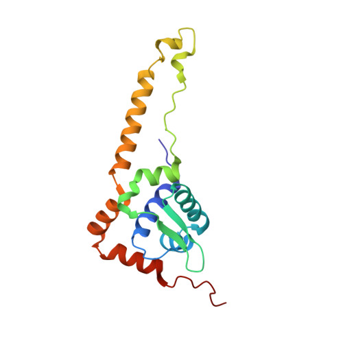
    The recently discovered Rrf2 family transcriptional regulator RsrR coordinates a [2Fe-2S] cluster. Remarkably, binding of the protein to RsrR-regulated promoter DNA sequences is switched on and off through the facile cycling of the [2Fe-2S] cluster between +2 and +1 states. Here, we report high resolution crystal structures of the RsrR dimer, revealing that the [2Fe-2S] cluster is asymmetrically coordinated across the RsrR monomer-monomer interface by two Cys residues from one subunit and His and Glu residues from the other. To our knowledge, this is the first example of a protein bound [Fe-S] cluster with three different amino acid side chains as ligands, and of Glu acting as ligand to a [2Fe-2S] cluster. Analyses of RsrR structures revealed a conformational change, centered on Trp9, which results in a significant shift in the DNA-binding helix-turn-helix region.


  • Organizational Affiliation

    Université Grenoble Alpes, CEA, CNRS , IBS , Metalloproteins Unit, F-38044 Grenoble , France.


Macromolecules
Find similar proteins by:  (by identity cutoff)  |  3D Structure
Entity ID: 1
MoleculeChains Sequence LengthOrganismDetailsImage
Rrf2 family transcriptional regulator
A, B, C, D
166Streptomyces venezuelae ATCC 10712Mutation(s): 0 
Gene Names: SVEN_6563
UniProt
Find proteins for F2RGC9 (Streptomyces venezuelae (strain ATCC 10712 / CBS 650.69 / DSM 40230 / JCM 4526 / NBRC 13096 / PD 04745))
Explore F2RGC9 
Go to UniProtKB:  F2RGC9
Entity Groups  
Sequence Clusters30% Identity50% Identity70% Identity90% Identity95% Identity100% Identity
UniProt GroupF2RGC9
Sequence Annotations
Expand
  • Reference Sequence
Small Molecules
Ligands 4 Unique
IDChains Name / Formula / InChI Key2D Diagram3D Interactions
MES
Query on MES

Download Ideal Coordinates CCD File 
F [auth A],
P [auth C],
X [auth D]
2-(N-MORPHOLINO)-ETHANESULFONIC ACID
C6 H13 N O4 S
SXGZJKUKBWWHRA-UHFFFAOYSA-N
FES
Query on FES

Download Ideal Coordinates CCD File 
E [auth A],
L [auth B],
O [auth C],
W [auth D]
FE2/S2 (INORGANIC) CLUSTER
Fe2 S2
NIXDOXVAJZFRNF-UHFFFAOYSA-N
GOL
Query on GOL

Download Ideal Coordinates CCD File 
G [auth A]
H [auth A]
I [auth A]
J [auth A]
K [auth A]
G [auth A],
H [auth A],
I [auth A],
J [auth A],
K [auth A],
M [auth B],
N [auth B],
Q [auth C],
R [auth C],
S [auth C],
T [auth C],
U [auth C],
Y [auth D],
Z [auth D]
GLYCEROL
C3 H8 O3
PEDCQBHIVMGVHV-UHFFFAOYSA-N
CL
Query on CL

Download Ideal Coordinates CCD File 
AA [auth D],
V [auth C]
CHLORIDE ION
Cl
VEXZGXHMUGYJMC-UHFFFAOYSA-M
Experimental Data & Validation

Experimental Data

  • Method: X-RAY DIFFRACTION
  • Resolution: 1.60 Å
  • R-Value Free: 0.190 
  • R-Value Work: 0.140 
  • R-Value Observed: 0.143 
  • Space Group: P 1 21 1
Unit Cell:
Length ( Å )Angle ( ˚ )
a = 36.29α = 90
b = 94.51β = 93.24
c = 99.94γ = 90
Software Package:
Software NamePurpose
XDSdata reduction
Aimlessdata scaling
REFMACrefinement
PDB_EXTRACTdata extraction
PHENIXphasing

Structure Validation

View Full Validation Report


Entry History 

Deposition Data

Revision History  (Full details and data files)

  • Version 1.0: 2019-01-30
    Type: Initial release
  • Version 1.1: 2019-02-13
    Changes: Data collection, Database references
  • Version 1.2: 2019-02-20
    Changes: Data collection, Database references