


The crystal structure of type II Dehydroquinase from Butyrivibrio crossotus DSM 2876
Lapthorn, A.J., Ner, L., Roszak, A.W.To be published.
Experimental Data Snapshot
wwPDB Validation 3D Report Full Report

Entity ID: 1 | |||||
|---|---|---|---|---|---|
| Molecule | Chains | Sequence Length | Organism | Details | Image |
| 3-dehydroquinate dehydratase | 145 | Acidithiobacillus caldus SM-1 | Mutation(s): 0 Gene Names: aroQ, Atc_2874 EC: 4.2.1.10 | 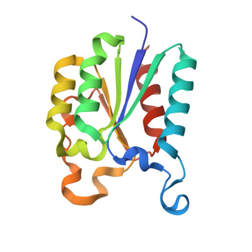 | |
UniProt | |||||
Find proteins for F9ZTU4 (Acidithiobacillus caldus (strain SM-1)) Explore F9ZTU4 Go to UniProtKB: F9ZTU4 | |||||
Entity Groups | |||||
| Sequence Clusters | 30% Identity50% Identity70% Identity90% Identity95% Identity100% Identity | ||||
| UniProt Group | F9ZTU4 | ||||
Sequence AnnotationsExpand | |||||
| |||||
| Ligands 2 Unique | |||||
|---|---|---|---|---|---|
| ID | Chains | Name / Formula / InChI Key | 2D Diagram | 3D Interactions | |
| EPE Query on EPE | D [auth A] | 4-(2-HYDROXYETHYL)-1-PIPERAZINE ETHANESULFONIC ACID C8 H18 N2 O4 S JKMHFZQWWAIEOD-UHFFFAOYSA-N |  | ||
| GOL Query on GOL | B [auth A], C [auth A] | GLYCEROL C3 H8 O3 PEDCQBHIVMGVHV-UHFFFAOYSA-N |  | ||
| Length ( Å ) | Angle ( ˚ ) |
|---|---|
| a = 188.458 | α = 90 |
| b = 188.458 | β = 90 |
| c = 188.458 | γ = 90 |
| Software Name | Purpose |
|---|---|
| REFMAC | refinement |
| XDS | data reduction |
| Aimless | data scaling |
| PHASER | phasing |
| PDB_EXTRACT | data extraction |
| Funding Organization | Location | Grant Number |
|---|---|---|
| Engineering and Physical Sciences Research Council | United Kingdom | EP/P00086X/1 |
RCSB PDB (citation) is hosted by
RCSB PDB is a member of the

