6HR1

Crystal structure of the YFPnano fusion protein


Experimental Data Snapshot

  • Method: X-RAY DIFFRACTION
  • Resolution: 1.90 Å
  • R-Value Free: 0.214 
  • R-Value Work: 0.181 
  • R-Value Observed: 0.182 

wwPDB Validation   3D Report Full Report


This is version 1.3 of the entry. See complete history


Literature

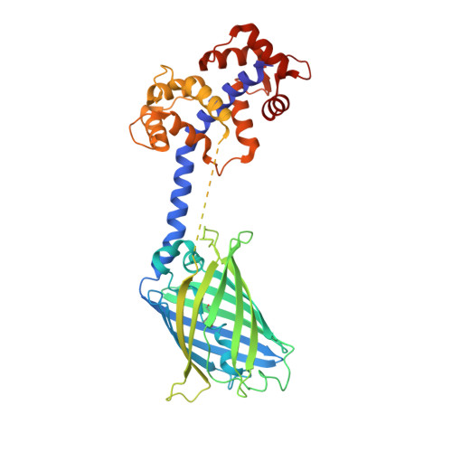
Chimeric single alpha-helical domains as rigid fusion protein connections for protein nanotechnology and structural biology.

Collu, G.Bierig, T.Krebs, A.S.Engilberge, S.Varma, N.Guixa-Gonzalez, R.Sharpe, T.Deupi, X.Olieric, V.Poghosyan, E.Benoit, R.M.

(2021) Structure 

  • DOI: https://doi.org/10.1016/j.str.2021.09.002
  • Primary Citation of Related Structures:  
    6HR1, 6XYR, 6YT3

  • PubMed Abstract: 

    Chimeric fusion proteins are essential tools for protein nanotechnology. Non-optimized protein-protein connections are usually flexible and therefore unsuitable as structural building blocks. Here we show that the ER/K motif, a single α-helical domain (SAH), can be seamlessly fused to terminal helices of proteins, forming an extended, partially free-standing rigid helix. This enables the connection of two domains at a defined distance and orientation. We designed three constructs termed YFPnano, T4Lnano, and MoStoNano. Analysis of experimentally determined structures and molecular dynamics simulations reveals a certain degree of plasticity in the connections that allows the adaptation to crystal contact opportunities. Our data show that SAHs can be stably integrated into designed structural elements, enabling new possibilities for protein nanotechnology, for example, to improve the exposure of epitopes on nanoparticles (structural vaccinology), to engineer crystal contacts with minimal impact on construct flexibility (for the study of protein dynamics), and to design novel biomaterials.


  • Organizational Affiliation

    Laboratory of Nanoscale Biology, Division of Biology and Chemistry, Paul Scherrer Institute, 5232 Villigen PSI, Switzerland; Department of Biology, ETH Zürich, 8093 Zürich, Switzerland.


Macromolecules
Find similar proteins by:  (by identity cutoff)  |  3D Structure
Entity ID: 1
MoleculeChains Sequence LengthOrganismDetailsImage
Myosin light chain kinase 2, skeletal/cardiac muscle,Unconventional myosin-X,Green fluorescent protein,Calmodulin-1
A, B
434Oryctolagus cuniculusBos taurusAequorea victoriaHomo sapiens
This entity is chimeric
Mutation(s): 2 
Gene Names: MYLK2MYO10GFPCALM1CALMCAMCAM1
EC: 2.7.11.18
UniProt & NIH Common Fund Data Resources
Find proteins for P42212 (Aequorea victoria)
Explore P42212 
Go to UniProtKB:  P42212
Find proteins for P0DP23 (Homo sapiens)
Explore P0DP23 
Go to UniProtKB:  P0DP23
PHAROS:  P0DP23
GTEx:  ENSG00000198668 
Find proteins for P79114 (Bos taurus)
Explore P79114 
Go to UniProtKB:  P79114
Find proteins for P07313 (Oryctolagus cuniculus)
Explore P07313 
Go to UniProtKB:  P07313
Entity Groups  
Sequence Clusters30% Identity50% Identity70% Identity90% Identity95% Identity100% Identity
UniProt GroupsP0DP23P42212P07313P79114
Sequence Annotations
Expand
  • Reference Sequence
Small Molecules
Ligands 5 Unique
IDChains Name / Formula / InChI Key2D Diagram3D Interactions
TLA
Query on TLA

Download Ideal Coordinates CCD File 
C [auth A]L(+)-TARTARIC ACID
C4 H6 O6
FEWJPZIEWOKRBE-JCYAYHJZSA-N
GOL
Query on GOL

Download Ideal Coordinates CCD File 
AA [auth B],
J [auth A],
K [auth A],
L [auth A],
Z [auth B]
GLYCEROL
C3 H8 O3
PEDCQBHIVMGVHV-UHFFFAOYSA-N
EDO
Query on EDO

Download Ideal Coordinates CCD File 
H [auth A]
I [auth A]
S [auth B]
T [auth B]
U [auth B]
H [auth A],
I [auth A],
S [auth B],
T [auth B],
U [auth B],
V [auth B],
W [auth B],
X [auth B],
Y [auth B]
1,2-ETHANEDIOL
C2 H6 O2
LYCAIKOWRPUZTN-UHFFFAOYSA-N
CA
Query on CA

Download Ideal Coordinates CCD File 
D [auth A]
E [auth A]
F [auth A]
G [auth A]
M [auth B]
D [auth A],
E [auth A],
F [auth A],
G [auth A],
M [auth B],
N [auth B],
O [auth B],
P [auth B]
CALCIUM ION
Ca
BHPQYMZQTOCNFJ-UHFFFAOYSA-N
NA
Query on NA

Download Ideal Coordinates CCD File 
Q [auth B],
R [auth B]
SODIUM ION
Na
FKNQFGJONOIPTF-UHFFFAOYSA-N
Modified Residues  1 Unique
IDChains TypeFormula2D DiagramParent
CR2
Query on CR2
A, B
L-PEPTIDE LINKINGC13 H13 N3 O4GLY, TYR, GLY
Experimental Data & Validation

Experimental Data

  • Method: X-RAY DIFFRACTION
  • Resolution: 1.90 Å
  • R-Value Free: 0.214 
  • R-Value Work: 0.181 
  • R-Value Observed: 0.182 
  • Space Group: P 1 21 1
Unit Cell:
Length ( Å )Angle ( ˚ )
a = 53.7α = 90
b = 117.8β = 99.9
c = 84.5γ = 90
Software Package:
Software NamePurpose
PHENIXrefinement
XDSdata reduction
XSCALEdata scaling
PHASERphasing
PDB_EXTRACTdata extraction

Structure Validation

View Full Validation Report


Entry History & Funding Information

Deposition Data

  • Released Date: 2020-04-08 
  • Deposition Author(s): Benoit, R.M.

Funding OrganizationLocationGrant Number
Switzerland--
Switzerland--

Revision History  (Full details and data files)

  • Version 1.0: 2020-04-08
    Type: Initial release
  • Version 1.1: 2020-10-21
    Changes: Database references, Derived calculations
  • Version 1.2: 2021-10-13
    Changes: Advisory, Data collection, Database references
  • Version 1.3: 2024-01-24
    Changes: Data collection, Database references, Refinement description