6HIT

The crystal structure of haemoglobin from Atlantic cod


Experimental Data Snapshot

  • Method: X-RAY DIFFRACTION
  • Resolution: 2.50 Å
  • R-Value Free: 0.301 
  • R-Value Work: 0.232 
  • R-Value Observed: 0.236 

wwPDB Validation   3D Report Full Report


Ligand Structure Quality Assessment 


This is version 1.1 of the entry. See complete history


Literature

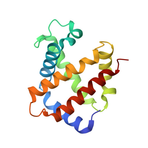
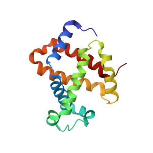
The crystal structure of haemoglobin from Atlantic cod.

Helland, R.Bjorkeng, E.K.Rothweiler, U.Sydnes, M.O.Pampanin, D.M.

(2019) Acta Crystallogr F Struct Biol Commun 75: 537-542

  • DOI: https://doi.org/10.1107/S2053230X1900904X
  • Primary Citation of Related Structures:  
    6HIT

  • PubMed Abstract: 

    The crystal structure of haemoglobin from Atlantic cod has been solved to 2.54 Å resolution. The structure consists of two tetramers in the crystallographic asymmetric unit. The structure of haemoglobin obtained from one individual cod suggests polymorphism in the tetrameric assembly.


  • Organizational Affiliation

    NorStruct, Department of Chemistry, Faculty of Science and Technology, UiT - The Arctic University of Norway, NO-9037 Tromsø, Norway.


Macromolecules
Find similar proteins by:  (by identity cutoff)  |  3D Structure
Entity ID: 1
MoleculeChains Sequence LengthOrganismDetailsImage
Hemoglobin alpha 2 chain
A, C, E, G
143Gadus morhuaMutation(s): 1 
UniProt
Find proteins for O42425 (Gadus morhua)
Explore O42425 
Go to UniProtKB:  O42425
Entity Groups  
Sequence Clusters30% Identity50% Identity70% Identity90% Identity95% Identity100% Identity
UniProt GroupO42425
Sequence Annotations
Expand
  • Reference Sequence
Find similar proteins by:  (by identity cutoff)  |  3D Structure
Entity ID: 2
MoleculeChains Sequence LengthOrganismDetailsImage
Hemoglobin beta 4 chain
B, D, F, H
145Gadus morhuaMutation(s): 0 
UniProt
Find proteins for B3F9D7 (Gadus morhua)
Explore B3F9D7 
Go to UniProtKB:  B3F9D7
Entity Groups  
Sequence Clusters30% Identity50% Identity70% Identity90% Identity95% Identity100% Identity
UniProt GroupB3F9D7
Sequence Annotations
Expand
  • Reference Sequence
Experimental Data & Validation

Experimental Data

  • Method: X-RAY DIFFRACTION
  • Resolution: 2.50 Å
  • R-Value Free: 0.301 
  • R-Value Work: 0.232 
  • R-Value Observed: 0.236 
  • Space Group: P 21 21 21
Unit Cell:
Length ( Å )Angle ( ˚ )
a = 62.691α = 90
b = 103.257β = 90
c = 199.352γ = 90
Software Package:
Software NamePurpose
REFMACrefinement
XDSdata reduction
Aimlessdata scaling
PHASERphasing
PDB_EXTRACTdata extraction

Structure Validation

View Full Validation Report


Ligand Structure Quality Assessment 


Entry History & Funding Information

Deposition Data


Funding OrganizationLocationGrant Number
Research Council of NorwayNorway229153
Research Council of NorwayNorway247732

Revision History  (Full details and data files)

  • Version 1.0: 2019-08-21
    Type: Initial release
  • Version 1.1: 2024-05-01
    Changes: Advisory, Data collection, Database references, Derived calculations, Refinement description