6H5L

Kuenenia stuttgartiensis reducing HAO-like protein complex Kustc0457/Kustc0458


Experimental Data Snapshot

  • Method: X-RAY DIFFRACTION
  • Resolution: 2.60 Å
  • R-Value Free: 0.234 
  • R-Value Work: 0.189 
  • R-Value Observed: 0.192 

wwPDB Validation   3D Report Full Report


Ligand Structure Quality Assessment 


This is version 1.1 of the entry. See complete history


Literature

A 60-heme reductase complex from an anammox bacterium shows an extended electron transfer pathway.

Dietl, A.Maalcke, W.J.Ferousi, C.Jetten, M.S.M.Kartal, B.Barends, T.R.M.

(2019) Acta Crystallogr D Struct Biol 75: 333-341

  • DOI: https://doi.org/10.1107/S2059798318017473
  • Primary Citation of Related Structures:  
    6H5L

  • PubMed Abstract: 

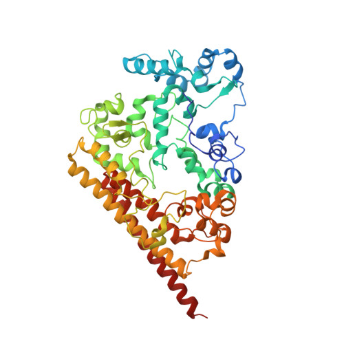
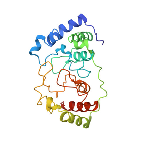
    The hydroxylamine oxidoreductase/hydrazine dehydrogenase (HAO/HDH) protein family constitutes an important group of octaheme cytochromes c (OCCs). The majority of these proteins form homotrimers, with their subunits being covalently attached to each other via a rare cross-link between the catalytic heme moiety and a conserved tyrosine residue in an adjacent subunit. This covalent cross-link has been proposed to modulate the active-site heme towards oxidative catalysis by distorting the heme plane. In this study, the crystal structure of a stable complex of an HAO homologue (KsHAOr) with its diheme cytochrome c redox partner (KsDH) from the anammox bacterium Kuenenia stuttgartiensis was determined. KsHAOr lacks the tyrosine cross-link and is therefore tuned to reductive catalysis. The molecular model of the KsHAOr-KsDH complex at 2.6 Å resolution shows a heterododecameric (α 6 β 6 ) assembly, which was also shown to be the oligomeric state in solution by analytical ultracentrifugation and multi-angle static light scattering. The 60-heme-containing protein complex reveals a unique extended electron transfer pathway and provides deeper insights into catalysis and electron transfer in reductive OCCs.


  • Organizational Affiliation

    Department of Biomolecular Mechanisms, Max Planck Institute for Medical Research, Jahnstrasse 29, 69120 Heidelberg, Germany.


Macromolecules
Find similar proteins by:  (by identity cutoff)  |  3D Structure
Entity ID: 1
MoleculeChains Sequence LengthOrganismDetailsImage
Similar to hydroxylamine oxidoreductase517Candidatus Kuenenia stuttgartiensisMutation(s): 0 
EC: 1.7.3.4
UniProt
Find proteins for A0A2C9CG41 (Kuenenia stuttgartiensis)
Explore A0A2C9CG41 
Go to UniProtKB:  A0A2C9CG41
Entity Groups  
Sequence Clusters30% Identity50% Identity70% Identity90% Identity95% Identity100% Identity
UniProt GroupA0A2C9CG41
Sequence Annotations
Expand
  • Reference Sequence
Find similar proteins by:  (by identity cutoff)  |  3D Structure
Entity ID: 2
MoleculeChains Sequence LengthOrganismDetailsImage
Conserved hypothetical cytochrome protein226Candidatus Kuenenia stuttgartiensisMutation(s): 0 
UniProt
Find proteins for Q1PVE1 (Kuenenia stuttgartiensis)
Explore Q1PVE1 
Go to UniProtKB:  Q1PVE1
Entity Groups  
Sequence Clusters30% Identity50% Identity70% Identity90% Identity95% Identity100% Identity
UniProt GroupQ1PVE1
Sequence Annotations
Expand
  • Reference Sequence
Experimental Data & Validation

Experimental Data

  • Method: X-RAY DIFFRACTION
  • Resolution: 2.60 Å
  • R-Value Free: 0.234 
  • R-Value Work: 0.189 
  • R-Value Observed: 0.192 
  • Space Group: H 3 2
Unit Cell:
Length ( Å )Angle ( ˚ )
a = 140.2α = 90
b = 140.2β = 90
c = 262.11γ = 120
Software Package:
Software NamePurpose
PHENIXrefinement
XDSdata reduction
XSCALEdata scaling
SHARPphasing

Structure Validation

View Full Validation Report


Ligand Structure Quality Assessment 


Entry History 

Deposition Data

Revision History  (Full details and data files)

  • Version 1.0: 2019-04-10
    Type: Initial release
  • Version 1.1: 2019-04-17
    Changes: Data collection, Database references