6H1Q

Proteus mirabilis Ambient Temperature Fimbriae adhesin AtfE


Experimental Data Snapshot

  • Method: X-RAY DIFFRACTION
  • Resolution: 1.58 Å
  • R-Value Free: 0.203 
  • R-Value Work: 0.179 
  • R-Value Observed: 0.180 

wwPDB Validation   3D Report Full Report


This is version 1.1 of the entry. See complete history


Literature

Structures of two fimbrial adhesins, AtfE and UcaD, from the uropathogen Proteus mirabilis.

Jiang, W.Ubhayasekera, W.Pearson, M.M.Knight, S.D.

(2018) Acta Crystallogr D Struct Biol 74: 1053-1062

  • DOI: https://doi.org/10.1107/S2059798318012391
  • Primary Citation of Related Structures:  
    6H1Q, 6H1X, 6H2L

  • PubMed Abstract: 

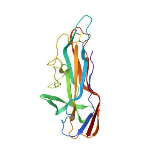
    The important uropathogen Proteus mirabilis encodes a record number of chaperone/usher-pathway adhesive fimbriae. Such fimbriae, which are used for adhesion to cell surfaces/tissues and for biofilm formation, are typically important virulence factors in bacterial pathogenesis. Here, the structures of the receptor-binding domains of the tip-located two-domain adhesins UcaD (1.5 Å resolution) and AtfE (1.58 Å resolution) from two P. mirabilis fimbriae (UCA/NAF and ATF) are presented. The structures of UcaD and AtfE are both similar to the F17G type of tip-located fimbrial receptor-binding domains, and the structures are very similar despite having only limited sequence similarity. These structures represent an important step towards a molecular-level understanding of P. mirabilis fimbrial adhesins and their roles in the complex pathogenesis of urinary-tract infections.


  • Organizational Affiliation

    Department of Cell and Molecular Biology, Uppsala University, Biomedical Center, PO Box 596, SE-751 24 Uppsala, Sweden.


Macromolecules
Find similar proteins by:  (by identity cutoff)  |  3D Structure
Entity ID: 1
MoleculeChains Sequence LengthOrganismDetailsImage
Fimbrial adhesin
A, B
193Proteus mirabilis HI4320Mutation(s): 0 
Gene Names: PMI2732
UniProt
Find proteins for B4EYH7 (Proteus mirabilis (strain HI4320))
Explore B4EYH7 
Go to UniProtKB:  B4EYH7
Entity Groups  
Sequence Clusters30% Identity50% Identity70% Identity90% Identity95% Identity100% Identity
UniProt GroupB4EYH7
Sequence Annotations
Expand
  • Reference Sequence
Experimental Data & Validation

Experimental Data

  • Method: X-RAY DIFFRACTION
  • Resolution: 1.58 Å
  • R-Value Free: 0.203 
  • R-Value Work: 0.179 
  • R-Value Observed: 0.180 
  • Space Group: C 1 2 1
Unit Cell:
Length ( Å )Angle ( ˚ )
a = 74.913α = 90
b = 103.793β = 95.829
c = 67.758γ = 90
Software Package:
Software NamePurpose
PHENIXrefinement
XDSdata reduction
Aimlessdata scaling
AutoSolphasing

Structure Validation

View Full Validation Report


Entry History & Funding Information

Deposition Data


Funding OrganizationLocationGrant Number
Swedish Research CouncilSweden--

Revision History  (Full details and data files)

  • Version 1.0: 2018-11-07
    Type: Initial release
  • Version 1.1: 2018-11-14
    Changes: Data collection, Database references