6GX9

Crystal structure of the TNPO3 - CPSF6 RSLD complex

  • Classification: NUCLEAR PROTEIN
  • Organism(s): Homo sapiens
  • Expression System: Escherichia coli
  • Mutation(s): No 

  • Deposited: 2018-06-26 Released: 2019-03-13 
  • Deposition Author(s): Cherepanov, P., Cook, N.
  • Funding Organization(s): National Institutes of Health/National Institute of Diabetes and Digestive and Kidney Disease (NIH/NIDDK)

Experimental Data Snapshot

  • Method: X-RAY DIFFRACTION
  • Resolution: 2.70 Å
  • R-Value Free: 0.248 
  • R-Value Work: 0.214 
  • R-Value Observed: 0.215 

wwPDB Validation   3D Report Full Report


Ligand Structure Quality Assessment 


This is version 1.4 of the entry. See complete history


Literature

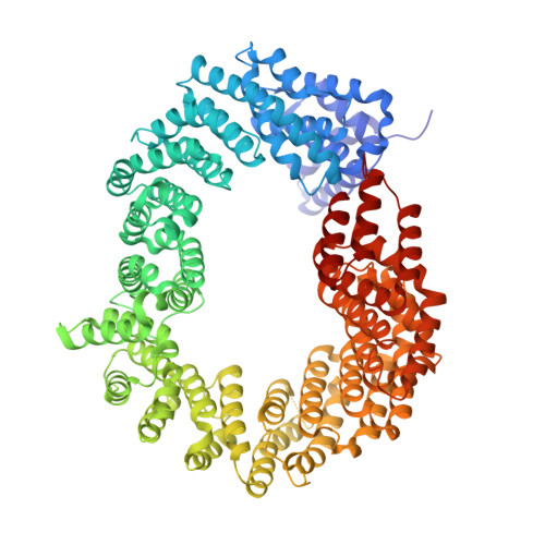
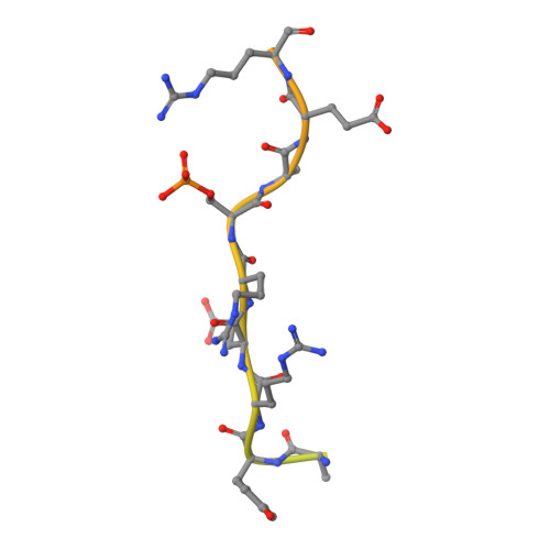
Differential role for phosphorylation in alternative polyadenylation function versus nuclear import of SR-like protein CPSF6.

Jang, S.Cook, N.J.Pye, V.E.Bedwell, G.J.Dudek, A.M.Singh, P.K.Cherepanov, P.Engelman, A.N.

(2019) Nucleic Acids Res 47: 4663-4683

  • DOI: https://doi.org/10.1093/nar/gkz206
  • Primary Citation of Related Structures:  
    6GX9

  • PubMed Abstract: 

    Cleavage factor I mammalian (CFIm) complex, composed of cleavage and polyadenylation specificity factor 5 (CPSF5) and serine/arginine-like protein CPSF6, regulates alternative polyadenylation (APA). Loss of CFIm function results in proximal polyadenylation site usage, shortening mRNA 3' untranslated regions (UTRs). Although CPSF6 plays additional roles in human disease, its nuclear translocation mechanism remains unresolved. Two β-karyopherins, transportin (TNPO) 1 and TNPO3, can bind CPSF6 in vitro, and we demonstrate here that while the TNPO1 binding site is dispensable for CPSF6 nuclear import, the arginine/serine (RS)-like domain (RSLD) that mediates TNPO3 binding is critical. The crystal structure of the RSLD-TNPO3 complex revealed potential CPSF6 interaction residues, which were confirmed to mediate TNPO3 binding and CPSF6 nuclear import. Both binding and nuclear import were independent of RSLD phosphorylation, though a hyperphosphorylated mimetic mutant failed to bind TNPO3 and mislocalized to the cell cytoplasm. Although hypophosphorylated CPSF6 largely supported normal polyadenylation site usage, a significant number of mRNAs harbored unnaturally extended 3' UTRs, similar to what is observed when other APA regulators, such as CFIIm component proteins, are depleted. Our results clarify the mechanism of CPSF6 nuclear import and highlight differential roles for RSLD phosphorylation in nuclear translocation versus regulation of APA.


  • Organizational Affiliation

    Department of Cancer Immunology and Virology, Dana-Farber Cancer Institute, Boston, MA 02215, USA.


Macromolecules
Find similar proteins by:  (by identity cutoff)  |  3D Structure
Entity ID: 1
MoleculeChains Sequence LengthOrganismDetailsImage
Transportin-3
A, B
923Homo sapiensMutation(s): 0 
Gene Names: TNPO3IPO12
UniProt & NIH Common Fund Data Resources
Find proteins for Q9Y5L0 (Homo sapiens)
Explore Q9Y5L0 
Go to UniProtKB:  Q9Y5L0
PHAROS:  Q9Y5L0
GTEx:  ENSG00000064419 
Entity Groups  
Sequence Clusters30% Identity50% Identity70% Identity90% Identity95% Identity100% Identity
UniProt GroupQ9Y5L0
Sequence Annotations
Expand
  • Reference Sequence
Find similar proteins by:  (by identity cutoff)  |  3D Structure
Entity ID: 2
MoleculeChains Sequence LengthOrganismDetailsImage
Cleavage and polyadenylation specificity factor subunit 6
C, D
70Homo sapiensMutation(s): 0 
Gene Names: CPSF6CFIM68
UniProt & NIH Common Fund Data Resources
Find proteins for Q16630 (Homo sapiens)
Explore Q16630 
Go to UniProtKB:  Q16630
PHAROS:  Q16630
GTEx:  ENSG00000111605 
Entity Groups  
Sequence Clusters30% Identity50% Identity70% Identity90% Identity95% Identity100% Identity
UniProt GroupQ16630
Sequence Annotations
Expand
  • Reference Sequence
Small Molecules
Modified Residues  1 Unique
IDChains TypeFormula2D DiagramParent
SEP
Query on SEP
C, D
L-PEPTIDE LINKINGC3 H8 N O6 PSER
Experimental Data & Validation

Experimental Data

  • Method: X-RAY DIFFRACTION
  • Resolution: 2.70 Å
  • R-Value Free: 0.248 
  • R-Value Work: 0.214 
  • R-Value Observed: 0.215 
  • Space Group: P 63 2 2
Unit Cell:
Length ( Å )Angle ( ˚ )
a = 200.335α = 90
b = 200.335β = 90
c = 234.807γ = 120
Software Package:
Software NamePurpose
PHENIXrefinement
MOSFLMdata reduction
SCALAdata scaling
PHASERphasing

Structure Validation

View Full Validation Report


Ligand Structure Quality Assessment 


Entry History & Funding Information

Deposition Data


Funding OrganizationLocationGrant Number
National Institutes of Health/National Institute of Diabetes and Digestive and Kidney Disease (NIH/NIDDK)United StatesGM082251

Revision History  (Full details and data files)

  • Version 1.0: 2019-03-13
    Type: Initial release
  • Version 1.1: 2019-04-10
    Changes: Data collection, Database references
  • Version 1.2: 2019-05-22
    Changes: Data collection, Database references
  • Version 1.3: 2022-03-30
    Changes: Author supporting evidence, Database references, Derived calculations
  • Version 1.4: 2024-01-17
    Changes: Data collection, Refinement description