6GTR

Structure of the AtaT Y144F mutant toxin bound to the C-terminus of the antitoxin AtaR and Acetyl-CoA


Experimental Data Snapshot

  • Method: X-RAY DIFFRACTION
  • Resolution: 2.99 Å
  • R-Value Free: 0.260 
  • R-Value Work: 0.193 
  • R-Value Observed: 0.196 

wwPDB Validation   3D Report Full Report


Ligand Structure Quality Assessment 


This is version 1.1 of the entry. See complete history


Literature

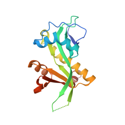
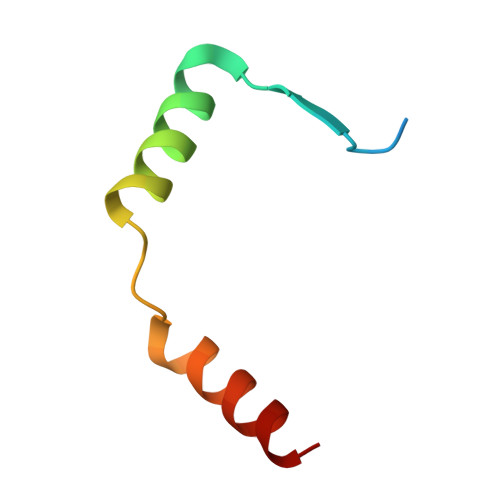
Mechanism of regulation and neutralization of the AtaR-AtaT toxin-antitoxin system.

Jurenas, D.Van Melderen, L.Garcia-Pino, A.

(2019) Nat Chem Biol 15: 285-294

  • DOI: https://doi.org/10.1038/s41589-018-0216-z
  • Primary Citation of Related Structures:  
    6GTO, 6GTP, 6GTQ, 6GTR, 6GTS

  • PubMed Abstract: 

    GCN5-related N-acetyl-transferase (GNAT)-like enzymes from toxin-antitoxin modules are strong inhibitors of protein synthesis. Here, we present the bases of the regulatory mechanisms of ataRT, a model GNAT-toxin-antitoxin module, from toxin synthesis to its action as a transcriptional de-repressor. We show the antitoxin (AtaR) traps the toxin (AtaT) in a pre-catalytic monomeric state and precludes the effective binding of ac-CoA and its target Met-transfer RNA fMet . In the repressor complex, AtaR intrinsically disordered region interacts with AtaT at two different sites, folding into different structures, that are involved in two separate functional roles, toxin neutralization and placing the DNA-binding domains of AtaR in a binding-compatible orientation. Our data suggests AtaR neutralizes AtaT as a monomer, right after its synthesis and only the toxin-antitoxin complex formed in this way is an active repressor. Once activated by dimerization, later neutralization of the toxin results in a toxin-antitoxin complex that is not able to repress transcription.


  • Organizational Affiliation

    Cellular and Molecular Microbiology, Department of Molecular Biology, Université Libre de Bruxelles, Gosselies, Belgium.


Macromolecules
Find similar proteins by:  (by identity cutoff)  |  3D Structure
Entity ID: 1
MoleculeChains Sequence LengthOrganismDetailsImage
N-acetyltransferase
A, B
178Escherichia coliMutation(s): 1 
Gene Names: BWP17_00640CVH05_12355
Entity Groups  
Sequence Clusters30% Identity50% Identity70% Identity90% Identity95% Identity100% Identity
Sequence Annotations
Expand
  • Reference Sequence
Find similar proteins by:  (by identity cutoff)  |  3D Structure
Entity ID: 2
MoleculeChains Sequence LengthOrganismDetailsImage
DUF1778 domain-containing protein
C, D
46Escherichia coliMutation(s): 0 
UniProt
Find proteins for J7QA90 (Escherichia coli)
Explore J7QA90 
Go to UniProtKB:  J7QA90
Entity Groups  
Sequence Clusters30% Identity50% Identity70% Identity90% Identity95% Identity100% Identity
UniProt GroupJ7QA90
Sequence Annotations
Expand
  • Reference Sequence
Experimental Data & Validation

Experimental Data

  • Method: X-RAY DIFFRACTION
  • Resolution: 2.99 Å
  • R-Value Free: 0.260 
  • R-Value Work: 0.193 
  • R-Value Observed: 0.196 
  • Space Group: P 31 2 1
Unit Cell:
Length ( Å )Angle ( ˚ )
a = 87.88α = 90
b = 87.88β = 90
c = 125.61γ = 120
Software Package:
Software NamePurpose
BUSTERrefinement
XDSdata reduction
XDSdata scaling
PHASERphasing

Structure Validation

View Full Validation Report


Ligand Structure Quality Assessment 


Entry History 

Deposition Data

Revision History  (Full details and data files)

  • Version 1.0: 2019-03-06
    Type: Initial release
  • Version 1.1: 2024-01-17
    Changes: Data collection, Database references, Refinement description