6GNX

Crystal structure of the MAJIN-TERB2 heterotetrameric complex - selenomethionine derivative


Experimental Data Snapshot

  • Method: X-RAY DIFFRACTION
  • Resolution: 2.90 Å
  • R-Value Free: 0.304 
  • R-Value Work: 0.254 
  • R-Value Observed: 0.259 

wwPDB Validation   3D Report Full Report


This is version 1.2 of the entry. See complete history


Literature

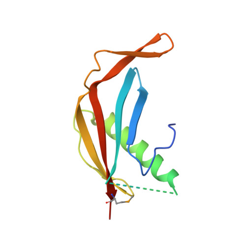
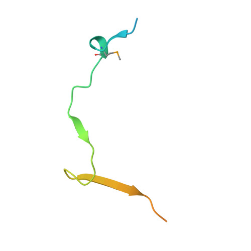
Structural basis of meiotic telomere attachment to the nuclear envelope by MAJIN-TERB2-TERB1.

Dunce, J.M.Milburn, A.E.Gurusaran, M.da Cruz, I.Sen, L.T.Benavente, R.Davies, O.R.

(2018) Nat Commun 9: 5355-5355

  • DOI: https://doi.org/10.1038/s41467-018-07794-7
  • Primary Citation of Related Structures:  
    6GNX, 6GNY

  • PubMed Abstract: 

    Meiotic chromosomes undergo rapid prophase movements, which are thought to facilitate the formation of inter-homologue recombination intermediates that underlie synapsis, crossing over and segregation. The meiotic telomere complex (MAJIN, TERB1, TERB2) tethers telomere ends to the nuclear envelope and transmits cytoskeletal forces via the LINC complex to drive these rapid movements. Here, we report the molecular architecture of the meiotic telomere complex through the crystal structure of MAJIN-TERB2, together with light and X-ray scattering studies of wider complexes. The MAJIN-TERB2 2:2 hetero-tetramer binds strongly to DNA and is tethered through long flexible linkers to the inner nuclear membrane and two TRF1-binding 1:1 TERB2-TERB1 complexes. Our complementary structured illumination microscopy studies and biochemical findings reveal a telomere attachment mechanism in which MAJIN-TERB2-TERB1 recruits telomere-bound TRF1, which is then displaced during pachytene, allowing MAJIN-TERB2-TERB1 to bind telomeric DNA and form a mature attachment plate.


  • Organizational Affiliation

    Institute for Cell and Molecular Biosciences, Faculty of Medical Sciences, Newcastle University, Framlington Place, Newcastle upon Tyne, NE2 4HH, UK.


Macromolecules
Find similar proteins by:  (by identity cutoff)  |  3D Structure
Entity ID: 1
MoleculeChains Sequence LengthOrganismDetailsImage
Membrane-anchored junction protein
A, C
114Homo sapiensMutation(s): 0 
Gene Names: MAJINC11orf85
UniProt & NIH Common Fund Data Resources
Find proteins for Q3KP22 (Homo sapiens)
Explore Q3KP22 
Go to UniProtKB:  Q3KP22
PHAROS:  Q3KP22
GTEx:  ENSG00000168070 
Entity Groups  
Sequence Clusters30% Identity50% Identity70% Identity90% Identity95% Identity100% Identity
UniProt GroupQ3KP22
Sequence Annotations
Expand
  • Reference Sequence
Find similar proteins by:  (by identity cutoff)  |  3D Structure
Entity ID: 2
MoleculeChains Sequence LengthOrganismDetailsImage
Telomere repeats-binding bouquet formation protein 2
B, D
55Homo sapiensMutation(s): 0 
Gene Names: TERB2C15orf43
UniProt & NIH Common Fund Data Resources
Find proteins for Q8NHR7 (Homo sapiens)
Explore Q8NHR7 
Go to UniProtKB:  Q8NHR7
PHAROS:  Q8NHR7
GTEx:  ENSG00000167014 
Entity Groups  
Sequence Clusters30% Identity50% Identity70% Identity90% Identity95% Identity100% Identity
UniProt GroupQ8NHR7
Sequence Annotations
Expand
  • Reference Sequence
Small Molecules
Modified Residues  1 Unique
IDChains TypeFormula2D DiagramParent
MSE
Query on MSE
A, C
L-PEPTIDE LINKINGC5 H11 N O2 SeMET
Experimental Data & Validation

Experimental Data

Unit Cell:
Length ( Å )Angle ( ˚ )
a = 59.88α = 90
b = 59.88β = 90
c = 159.93γ = 120
Software Package:
Software NamePurpose
PHENIXrefinement
XDSdata reduction
XSCALEdata scaling
PHENIXphasing

Structure Validation

View Full Validation Report


Entry History & Funding Information

Deposition Data


Funding OrganizationLocationGrant Number
Wellcome TrustUnited Kingdom104158/Z/14/Z
Royal SocietyUnited KingdomRG170118

Revision History  (Full details and data files)

  • Version 1.0: 2018-12-12
    Type: Initial release
  • Version 1.1: 2019-01-02
    Changes: Data collection, Database references
  • Version 1.2: 2020-04-22
    Changes: Database references