6GIE

Crystal structure of the Acinetobacter baumannii outer membrane protein Omp33


Experimental Data Snapshot

  • Method: X-RAY DIFFRACTION
  • Resolution: 2.10 Å
  • R-Value Free: 0.266 
  • R-Value Work: 0.213 
  • R-Value Observed: 0.216 

wwPDB Validation   3D Report Full Report


Ligand Structure Quality Assessment 


This is version 1.0 of the entry. See complete history


Literature

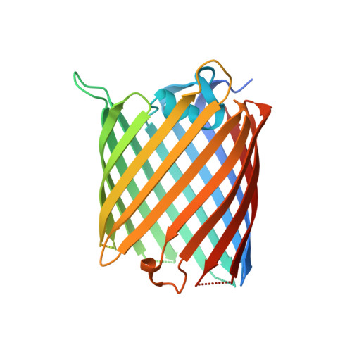
Crystal structure of the Acinetobacter baumannii outer membrane protein Omp33.

Abellon-Ruiz, J.Zahn, M.Basle, A.van den Berg, B.

(2018) Acta Crystallogr D Struct Biol 74: 852-860

  • DOI: https://doi.org/10.1107/S205979831800904X
  • Primary Citation of Related Structures:  
    6GIE

  • PubMed Abstract: 

    Acinetobacter baumannii is becoming a major threat to human health due to its multidrug resistance. This is owing in a large part to the low permeability of its outer membrane (OM), which prevents high internal antibiotic concentrations and makes antibiotic-resistance mechanisms more effective. To exploit OM channels as potential delivery vehicles for future antibiotics, structural information is required. One abundant OM protein in A. baumannii is Omp33. This protein has been reported to be important for the in vivo fitness and virulence of A. baumannii, but its structure is not known. Here, the X-ray crystal structure of Omp33 is reported at a resolution of 2.1 Å. Omp33 has a 14-β-stranded barrel without stable extracellular loop constrictions. Instead, an extended and unusual periplasmic turn connecting β-strands 2 and 3 is present, which folds into the pore lumen and completely blocks the aqueous channel. The Omp33 structure helps in understanding how A. baumannii OM proteins contribute to the low permeability of the cell envelope of this bacterium and suggests that Omp33 might function as a gated channel.


  • Organizational Affiliation

    Institute for Cell and Molecular Biosciences, The Medical School, Newcastle University, Newcastle upon Tyne NE2 4HH, England.


Macromolecules
Find similar proteins by:  (by identity cutoff)  |  3D Structure
Entity ID: 1
MoleculeChains Sequence LengthOrganismDetailsImage
33-36 kDa outer membrane protein296Acinetobacter baumanniiMutation(s): 0 
Gene Names: carOABUW_0187B4R90_18580CBI29_03686CHQ89_12430CV949_17495CV951_18190CV952_18425CV954_00850
Membrane Entity: Yes 
UniProt
Find proteins for A0A081GU02 (Acinetobacter baumannii)
Explore A0A081GU02 
Go to UniProtKB:  A0A081GU02
Entity Groups  
Sequence Clusters30% Identity50% Identity70% Identity90% Identity95% Identity100% Identity
UniProt GroupA0A081GU02
Sequence Annotations
Expand
  • Reference Sequence
Experimental Data & Validation

Experimental Data

  • Method: X-RAY DIFFRACTION
  • Resolution: 2.10 Å
  • R-Value Free: 0.266 
  • R-Value Work: 0.213 
  • R-Value Observed: 0.216 
  • Space Group: P 21 21 2
Unit Cell:
Length ( Å )Angle ( ˚ )
a = 69.68α = 90
b = 73.74β = 90
c = 53.03γ = 90
Software Package:
Software NamePurpose
REFMACrefinement
XDSdata reduction
Aimlessdata scaling
PHASERphasing
Cootmodel building

Structure Validation

View Full Validation Report


Ligand Structure Quality Assessment 


Entry History & Funding Information

Deposition Data


Funding OrganizationLocationGrant Number
European UnionUnited Kingdom115525

Revision History  (Full details and data files)

  • Version 1.0: 2018-09-19
    Type: Initial release