


Structure of the H1 domain of human KCTD8
Pinkas, D.M.To be published.
Experimental Data Snapshot
wwPDB Validation 3D Report Full Report

Entity ID: 1 | |||||
|---|---|---|---|---|---|
| Molecule | Chains | Sequence Length | Organism | Details | Image |
| BTB/POZ domain-containing protein KCTD8 | 124 | Homo sapiens | Mutation(s): 0 Gene Names: KCTD8 | 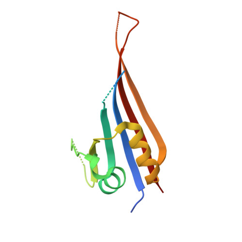 | |
UniProt & NIH Common Fund Data Resources | |||||
Find proteins for Q6ZWB6 (Homo sapiens) Explore Q6ZWB6 Go to UniProtKB: Q6ZWB6 | |||||
PHAROS: Q6ZWB6 GTEx: ENSG00000183783 | |||||
Entity Groups | |||||
| Sequence Clusters | 30% Identity50% Identity70% Identity90% Identity95% Identity100% Identity | ||||
| UniProt Group | Q6ZWB6 | ||||
Sequence AnnotationsExpand | |||||
| |||||
| Length ( Å ) | Angle ( ˚ ) |
|---|---|
| a = 92.117 | α = 90 |
| b = 92.117 | β = 90 |
| c = 219.355 | γ = 120 |
| Software Name | Purpose |
|---|---|
| PHENIX | refinement |
| xia2 | data reduction |
| xia2 | data scaling |
| PHASER | phasing |
RCSB PDB (citation) is hosted by
RCSB PDB is a member of the

