


6G1U
Crystal structure of Torpedo Californica acetylcholinesterase in complex with 9-Amino-6-chloro-1,2,3,4-tetrahydro-10-methylacridin-10-ium
- PDB DOI: https://doi.org/10.2210/pdb6G1U/pdb
- Entry: 6G1U supersedes: 6FOT
- Classification: HYDROLASE
- Organism(s): Tetronarce californica
- Mutation(s): No
- Deposited: 2018-03-22 Released: 2018-04-04
Experimental Data Snapshot
- Method: X-RAY DIFFRACTION
- Resolution: 1.79 Å
- R-Value Free: 0.213
- R-Value Work: 0.180
- R-Value Observed: 0.181

This is version 2.2 of the entry. See complete history.

Macromolecules
Find similar proteins by:
(by identity cutoff) | 3D Structure
Entity ID: 1 | |||||
|---|---|---|---|---|---|
| Molecule | Chains | Sequence Length | Organism | Details | Image |
| Acetylcholinesterase | 565 | Tetronarce californica | Mutation(s): 0 EC: 3.1.1.7 | 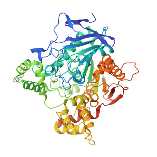 | |
UniProt | |||||
Find proteins for P04058 (Tetronarce californica) Explore P04058 Go to UniProtKB: P04058 | |||||
Entity Groups | |||||
| Sequence Clusters | 30% Identity50% Identity70% Identity90% Identity95% Identity100% Identity | ||||
| UniProt Group | P04058 | ||||
Sequence AnnotationsExpand | |||||
| |||||
Small Molecules
| Ligands 4 Unique | |||||
|---|---|---|---|---|---|
| ID | Chains | Name / Formula / InChI Key | 2D Diagram | 3D Interactions | |
| E1K Query on E1K | I [auth A] J [auth A] K [auth A] L [auth A] U [auth B] | 6-chloranyl-10-methyl-1,2,3,4-tetrahydroacridin-10-ium-9-amine C14 H16 Cl N2 NNLSQYGKFPZYHI-UHFFFAOYSA-O |  | ||
| NAG Query on NAG | AA [auth B] C [auth A] D [auth A] E [auth A] Q [auth B] | 2-acetamido-2-deoxy-beta-D-glucopyranose C8 H15 N O6 OVRNDRQMDRJTHS-FMDGEEDCSA-N |  | ||
| PEG Query on PEG | F [auth A] G [auth A] H [auth A] O [auth A] P [auth A] | DI(HYDROXYETHYL)ETHER C4 H10 O3 MTHSVFCYNBDYFN-UHFFFAOYSA-N |  | ||
| CL Query on CL | M [auth A], N [auth A], Y [auth B] | CHLORIDE ION Cl VEXZGXHMUGYJMC-UHFFFAOYSA-M |  | ||
Experimental Data & Validation
Experimental Data
- Method: X-RAY DIFFRACTION
- Resolution: 1.79 Å
- R-Value Free: 0.213
- R-Value Work: 0.180
- R-Value Observed: 0.181
- Space Group: P 21 21 21
Unit Cell:
| Length ( Å ) | Angle ( ˚ ) |
|---|---|
| a = 91.431 | α = 90 |
| b = 106.6 | β = 90 |
| c = 150.533 | γ = 90 |
| Software Name | Purpose |
|---|---|
| PHENIX | refinement |
| XDS | data reduction |
| XSCALE | data scaling |
| PHASER | phasing |
Entry History
Deposition Data
- Released Date: 2018-04-04 Deposition Author(s): Coquelle, N., Colletier, J.P.
- This entry supersedes: 6FOT
Revision History (Full details and data files)
- Version 1.0: 2018-04-04
Type: Initial release - Version 2.0: 2019-05-01
Changes: Advisory, Atomic model, Data collection, Database references, Derived calculations, Polymer sequence, Source and taxonomy, Structure summary - Version 2.1: 2020-07-29
Type: Remediation
Reason: Carbohydrate remediation
Changes: Data collection, Derived calculations, Structure summary - Version 2.2: 2024-01-17
Changes: Data collection, Database references, Refinement description, Structure summary
