


Crystal Structure of a radical SAM methyltransferase from Sphaerobacter thermophilus
Shaw, J.M., Hinchliffe, P., Spencer, J.To be published.
Experimental Data Snapshot
Entity ID: 1 | |||||
|---|---|---|---|---|---|
| Molecule | Chains | Sequence Length | Organism | Details | Image |
| Probable dual-specificity RNA methyltransferase RlmN | 366 | Sphaerobacter thermophilus DSM 20745 | Mutation(s): 0 Gene Names: rlmN, Sthe_1821 EC: 2.1.1.192 | 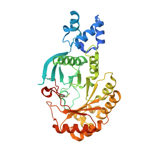 | |
UniProt | |||||
Find proteins for D1C4T7 (Sphaerobacter thermophilus (strain DSM 20745 / S 6022)) Explore D1C4T7 Go to UniProtKB: D1C4T7 | |||||
Entity Groups | |||||
| Sequence Clusters | 30% Identity50% Identity70% Identity90% Identity95% Identity100% Identity | ||||
| UniProt Group | D1C4T7 | ||||
Sequence AnnotationsExpand | |||||
| |||||
| Ligands 4 Unique | |||||
|---|---|---|---|---|---|
| ID | Chains | Name / Formula / InChI Key | 2D Diagram | 3D Interactions | |
| SAH Query on SAH | D [auth A], H [auth B] | S-ADENOSYL-L-HOMOCYSTEINE C14 H20 N6 O5 S ZJUKTBDSGOFHSH-WFMPWKQPSA-N |  | ||
| SF4 Query on SF4 | C [auth A], G [auth B] | IRON/SULFUR CLUSTER Fe4 S4 LJBDFODJNLIPKO-UHFFFAOYSA-N |  | ||
| GOL Query on GOL | E [auth A] | GLYCEROL C3 H8 O3 PEDCQBHIVMGVHV-UHFFFAOYSA-N |  | ||
| BR Query on BR | F [auth A], I [auth B] | BROMIDE ION Br CPELXLSAUQHCOX-UHFFFAOYSA-M |  | ||
| Modified Residues 1 Unique | |||||
|---|---|---|---|---|---|
| ID | Chains | Type | Formula | 2D Diagram | Parent |
| SMC Query on SMC | A, B | L-PEPTIDE LINKING | C4 H9 N O2 S |  | CYS |
| Length ( Å ) | Angle ( ˚ ) |
|---|---|
| a = 131.84 | α = 90 |
| b = 53.14 | β = 90 |
| c = 113.57 | γ = 90 |
| Software Name | Purpose |
|---|---|
| PHENIX | refinement |
| xia2 | data reduction |
| xia2 | data scaling |
| AutoSol | phasing |
| Funding Organization | Location | Grant Number |
|---|---|---|
| Biotechnology and Biological Sciences Research Council | United Kingdom | BB/J017906/1 |
| Biotechnology and Biological Sciences Research Council | United Kingdom | BB/M012107/1 |
RCSB PDB (citation) is hosted by
RCSB PDB is a member of the

