6FVQ

The active form of a pentameric ion channel (sTeLIC) gated by alkaline pH - R86A


Experimental Data Snapshot

  • Method: X-RAY DIFFRACTION
  • Resolution: 3.30 Å
  • R-Value Free: 0.217 
  • R-Value Work: 0.204 
  • R-Value Observed: 0.205 

wwPDB Validation   3D Report Full Report


Ligand Structure Quality Assessment 


This is version 1.2 of the entry. See complete history


Literature

Crystal structures of a pentameric ion channel gated by alkaline pH show a widely open pore and identify a cavity for modulation.

Hu, H.Nemecz, A.Van Renterghem, C.Fourati, Z.Sauguet, L.Corringer, P.J.Delarue, M.

(2018) Proc Natl Acad Sci U S A 115: E3959-E3968

  • DOI: https://doi.org/10.1073/pnas.1717700115
  • Primary Citation of Related Structures:  
    6FL9, 6FLI, 6FVQ, 6FVR, 6FVS

  • PubMed Abstract: 

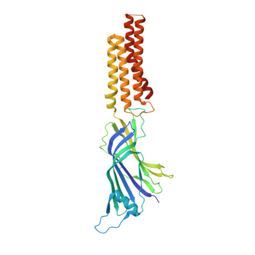
    Pentameric ligand-gated ion channels (pLGICs) constitute a widespread class of ion channels, present in archaea, bacteria, and eukaryotes. Upon binding of their agonists in the extracellular domain, the transmembrane pore opens, allowing ions to go through, via a gating mechanism that can be modulated by a number of drugs. Even though high-resolution structural information on pLGICs has increased in a spectacular way in recent years, both in bacterial and in eukaryotic systems, the structure of the open channel conformation of some intensively studied receptors whose structures are known in a nonactive (closed) form, such as Erwinia chrysanthemi pLGIC (ELIC), is still lacking. Here we describe a gammaproteobacterial pLGIC from an endo-symbiont of Tevnia jerichonana (sTeLIC), whose sequence is closely related to the pLGIC from ELIC with 28% identity. We provide an X-ray crystallographic structure at 2.3 Å in an active conformation, where the pore is found to be more open than any current conformation found for pLGICs. In addition, two charged restriction rings are present in the vestibule. Functional characterization shows sTeLIC to be a cationic channel activated at alkaline pH. It is inhibited by divalent cations, but not by quaternary ammonium ions, such as tetramethylammonium. Additionally, we found that sTeLIC is allosterically potentiated by aromatic amino acids Phe and Trp, as well as their derivatives, such as 4-bromo-cinnamate, whose cocrystal structure reveals a vestibular binding site equivalent to, but more deeply buried than, the one already described for benzodiazepines in ELIC.


  • Organizational Affiliation

    Unité Dynamique Structurale des Macromolécules, Institut Pasteur, UMR 3528, CNRS, 75015 Paris, France.


Macromolecules
Find similar proteins by:  (by identity cutoff)  |  3D Structure
Entity ID: 1
MoleculeChains Sequence LengthOrganismDetailsImage
Cys-loop ligand-gated ion channel
A, B, C, D, E
320endosymbiont of Tevnia jerichonana (vent Tica)Mutation(s): 1 
Gene Names: TevJSym_bc00020
Membrane Entity: Yes 
UniProt
Find proteins for G2FID1 (endosymbiont of Tevnia jerichonana)
Explore G2FID1 
Go to UniProtKB:  G2FID1
Entity Groups  
Sequence Clusters30% Identity50% Identity70% Identity90% Identity95% Identity100% Identity
UniProt GroupG2FID1
Sequence Annotations
Expand
  • Reference Sequence
Small Molecules
Ligands 2 Unique
IDChains Name / Formula / InChI Key2D Diagram3D Interactions
BNG
Query on BNG

Download Ideal Coordinates CCD File 
H [auth A],
I [auth A],
K [auth B],
M [auth C],
O [auth D]
nonyl beta-D-glucopyranoside
C15 H30 O6
QFAPUKLCALRPLH-UXXRCYHCSA-N
PG4
Query on PG4

Download Ideal Coordinates CCD File 
F [auth A],
G [auth A],
J [auth B],
L [auth C],
N [auth D]
TETRAETHYLENE GLYCOL
C8 H18 O5
UWHCKJMYHZGTIT-UHFFFAOYSA-N
Experimental Data & Validation

Experimental Data

  • Method: X-RAY DIFFRACTION
  • Resolution: 3.30 Å
  • R-Value Free: 0.217 
  • R-Value Work: 0.204 
  • R-Value Observed: 0.205 
  • Space Group: C 1 2 1
Unit Cell:
Length ( Å )Angle ( ˚ )
a = 216.237α = 90
b = 112.475β = 113.8
c = 144.461γ = 90
Software Package:
Software NamePurpose
BUSTERrefinement
XDSdata reduction
XDSdata scaling
PHASERphasing

Structure Validation

View Full Validation Report


Ligand Structure Quality Assessment 


Entry History 

Deposition Data

Revision History  (Full details and data files)

  • Version 1.0: 2018-05-16
    Type: Initial release
  • Version 1.1: 2019-06-12
    Changes: Data collection, Structure summary
  • Version 1.2: 2020-07-29
    Type: Remediation
    Reason: Carbohydrate remediation
    Changes: Data collection, Derived calculations, Structure summary