6FVN

DNA polymerase sliding clamp from Mycobacterium tuberculosis with bound P7 peptide


Experimental Data Snapshot

  • Method: X-RAY DIFFRACTION
  • Resolution: 3.14 Å
  • R-Value Free: 0.291 
  • R-Value Work: 0.256 
  • R-Value Observed: 0.258 

wwPDB Validation   3D Report Full Report


This is version 1.1 of the entry. See complete history


Literature

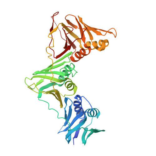
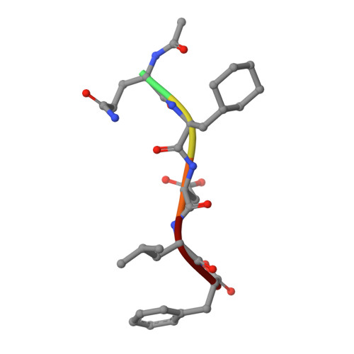
Peptide Interactions on Bacterial Sliding Clamps.

Andre, C.Martiel, I.Wolff, P.Landolfo, M.Lorber, B.Silva da Veiga, C.Dejaegere, A.Dumas, P.Guichard, G.Olieric, V.Wagner, J.G.Burnouf, D.Y.

(2019) ACS Infect Dis 

  • DOI: https://doi.org/10.1021/acsinfecdis.9b00089
  • Primary Citation of Related Structures:  
    6FVL, 6FVM, 6FVN, 6FVO

  • PubMed Abstract: 

    Bacterial sliding clamps control the access of DNA polymerases to the replication fork and are appealing targets for antibacterial drug development. It is therefore essential to decipher the polymerase-clamp binding mode across various bacterial species. Here, two residues of the E. coli clamp binding pocket, Ec S 346 and Ec M 362 , and their cognate residues in M. tuberculosis and B. subtilis clamps, were mutated. The effects of these mutations on the interaction of a model peptide with these variant clamps were evaluated by thermodynamic, molecular dynamics, X-rays crystallography, and biochemical analyses. Ec M 362 and corresponding residues in Gram positive clamps occupy a strategic position where a mobile residue is essential for an efficient peptide interaction. Ec S 346 has a more subtle function that modulates the pocket folding dynamics, while the equivalent residue in B. subtilis is essential for polymerase activity and might therefore be a Gram positive-specific molecular marker. Finally, the peptide binds through an induced-fit process to Gram negative and positive pockets, but the complex stability varies according to a pocket-specific network of interactions.


  • Organizational Affiliation

    Institut Européen de Chimie et Biologie , Université de Bordeaux-CNRS UMR 5248, CBMN , 2, rue Robert Escarpit , 33607 Pessac , France.


Macromolecules
Find similar proteins by:  (by identity cutoff)  |  3D Structure
Entity ID: 1
MoleculeChains Sequence LengthOrganismDetailsImage
Beta sliding clampA,
C [auth B],
E [auth C],
G [auth D]
402Mycobacterium tuberculosis CDC1551Mutation(s): 0 
Gene Names: dnaNMT0002
UniProt
Find proteins for P9WNU1 (Mycobacterium tuberculosis (strain ATCC 25618 / H37Rv))
Explore P9WNU1 
Go to UniProtKB:  P9WNU1
Entity Groups  
Sequence Clusters30% Identity50% Identity70% Identity90% Identity95% Identity100% Identity
UniProt GroupP9WNU1
Sequence Annotations
Expand
  • Reference Sequence

Find similar proteins by:  Sequence   |   3D Structure  

Entity ID: 2
MoleculeChains Sequence LengthOrganismDetailsImage
P7 peptideB [auth J],
D [auth K],
F [auth H],
H [auth I]
6synthetic constructMutation(s): 0 
Sequence Annotations
Expand
  • Reference Sequence
Small Molecules
Modified Residues  1 Unique
IDChains TypeFormula2D DiagramParent
ALC
Query on ALC
B [auth J],
D [auth K],
F [auth H],
H [auth I]
L-PEPTIDE LINKINGC9 H17 N O2ALA
Experimental Data & Validation

Experimental Data

  • Method: X-RAY DIFFRACTION
  • Resolution: 3.14 Å
  • R-Value Free: 0.291 
  • R-Value Work: 0.256 
  • R-Value Observed: 0.258 
  • Space Group: P 21 21 21
Unit Cell:
Length ( Å )Angle ( ˚ )
a = 79.447α = 90
b = 126.761β = 90
c = 171.02γ = 90
Software Package:
Software NamePurpose
XDSdata reduction
XSCALEdata scaling
STARANISOdata scaling
MOLREPphasing
BUSTERrefinement
PHENIXrefinement
PDB_EXTRACTdata extraction

Structure Validation

View Full Validation Report


Entry History & Funding Information

Deposition Data


Funding OrganizationLocationGrant Number
IMMIFrance2014013

Revision History  (Full details and data files)

  • Version 1.0: 2019-04-10
    Type: Initial release
  • Version 1.1: 2024-01-17
    Changes: Data collection, Database references, Refinement description