6FUE

Periplasmic coiled coil domain of the FapF amyloid transporter


Experimental Data Snapshot

  • Method: X-RAY DIFFRACTION
  • Resolution: 1.78 Å
  • R-Value Free: 0.248 
  • R-Value Work: 0.207 
  • R-Value Observed: 0.209 

wwPDB Validation   3D Report Full Report


This is version 1.1 of the entry. See complete history


Literature

The FapF Amyloid Secretion Transporter Possesses an Atypical Asymmetric Coiled Coil.

Rouse, S.L.Stylianou, F.Wu, H.Y.G.Berry, J.L.Sewell, L.Morgan, R.M.L.Sauerwein, A.C.Matthews, S.

(2018) J Mol Biol 430: 3863-3871

  • DOI: https://doi.org/10.1016/j.jmb.2018.06.007
  • Primary Citation of Related Structures:  
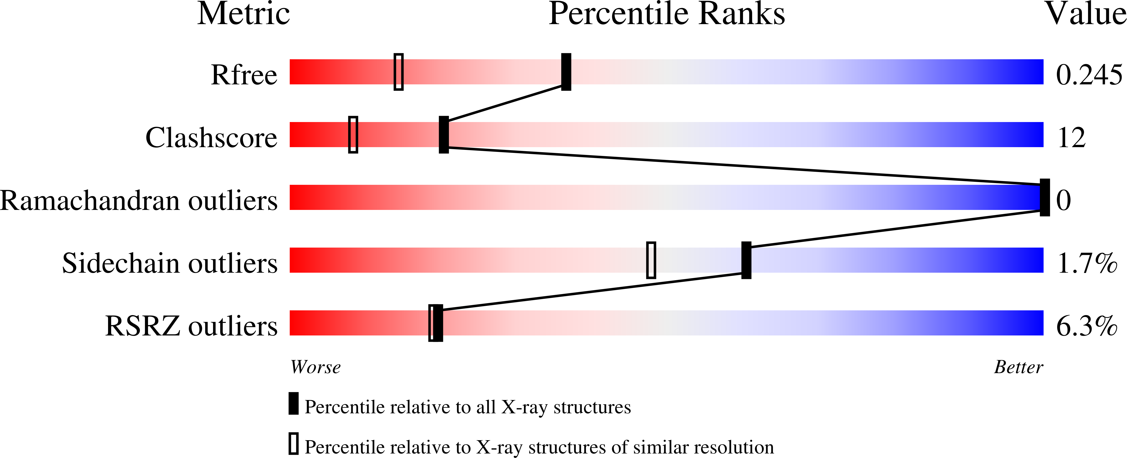
    6FUE

  • PubMed Abstract: 

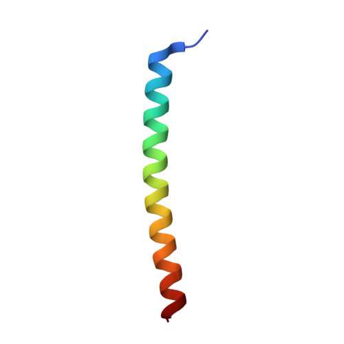
    Gram-negative bacteria possess specialized biogenesis machineries that facilitate the export of amyloid subunits, the fibers of which are key components of their biofilm matrix. The secretion of bacterial functional amyloid requires a specialized outer-membrane protein channel through which unfolded amyloid substrates are translocated. We previously reported the crystal structure of the membrane-spanning domain of the amyloid subunit transporter FapF from Pseudomonas. However, the structure of the periplasmic domain, which is essential for amyloid transport, is yet to be determined. Here, we present the crystal structure of the N-terminal periplasmic domain at 1.8-Å resolution. This domain forms a novel asymmetric trimeric coiled coil that possesses a single buried tyrosine residue as well as an extensive hydrogen-bonding network within a glutamine layer. This new structural insight allows us to understand this newly described functional amyloid secretion system in greater detail.


  • Organizational Affiliation

    Department of Life Sciences, Imperial College London, South Kensington Campus, London SW7 2AZ, UK.


Macromolecules
Find similar proteins by:  (by identity cutoff)  |  3D Structure
Entity ID: 1
MoleculeChains Sequence LengthOrganismDetailsImage
FapF
A, B, C, D, E
A, B, C, D, E, F
38Pseudomonas sp. UK4Mutation(s): 0 
UniProt
Find proteins for C4IN73 (Pseudomonas sp. UK4)
Explore C4IN73 
Go to UniProtKB:  C4IN73
Entity Groups  
Sequence Clusters30% Identity50% Identity70% Identity90% Identity95% Identity100% Identity
UniProt GroupC4IN73
Sequence Annotations
Expand
  • Reference Sequence
Small Molecules
Ligands 3 Unique
IDChains Name / Formula / InChI Key2D Diagram3D Interactions
ZN
Query on ZN

Download Ideal Coordinates CCD File 
G [auth A]
H [auth A]
I [auth A]
K [auth B]
Q [auth D]
G [auth A],
H [auth A],
I [auth A],
K [auth B],
Q [auth D],
S [auth E],
T [auth E],
U [auth F],
V [auth F],
W [auth F],
X [auth F]
ZINC ION
Zn
PTFCDOFLOPIGGS-UHFFFAOYSA-N
IPA
Query on IPA

Download Ideal Coordinates CCD File 
P [auth C]ISOPROPYL ALCOHOL
C3 H8 O
KFZMGEQAYNKOFK-UHFFFAOYSA-N
NA
Query on NA

Download Ideal Coordinates CCD File 
J [auth A]
L [auth B]
M [auth B]
N [auth B]
O [auth B]
J [auth A],
L [auth B],
M [auth B],
N [auth B],
O [auth B],
R [auth D]
SODIUM ION
Na
FKNQFGJONOIPTF-UHFFFAOYSA-N
Experimental Data & Validation

Experimental Data

  • Method: X-RAY DIFFRACTION
  • Resolution: 1.78 Å
  • R-Value Free: 0.248 
  • R-Value Work: 0.207 
  • R-Value Observed: 0.209 
  • Space Group: P 1
Unit Cell:
Length ( Å )Angle ( ˚ )
a = 37.59α = 90.24
b = 37.7β = 108.95
c = 57.34γ = 119.89
Software Package:
Software NamePurpose
REFMACrefinement
XDSdata reduction
SCALAdata scaling
MoRDaphasing

Structure Validation

View Full Validation Report


Entry History & Funding Information

Deposition Data


Funding OrganizationLocationGrant Number
Wellcome TrustUnited Kingdom--

Revision History  (Full details and data files)

  • Version 1.0: 2018-06-20
    Type: Initial release
  • Version 1.1: 2018-10-10
    Changes: Data collection, Database references, Structure summary