6FQB

MurT/GatD peptidoglycan amidotransferase complex from Streptococcus pneumoniae R6


Experimental Data Snapshot

  • Method: X-RAY DIFFRACTION
  • Resolution: 3.00 Å
  • R-Value Free: 0.228 
  • R-Value Work: 0.189 
  • R-Value Observed: 0.193 

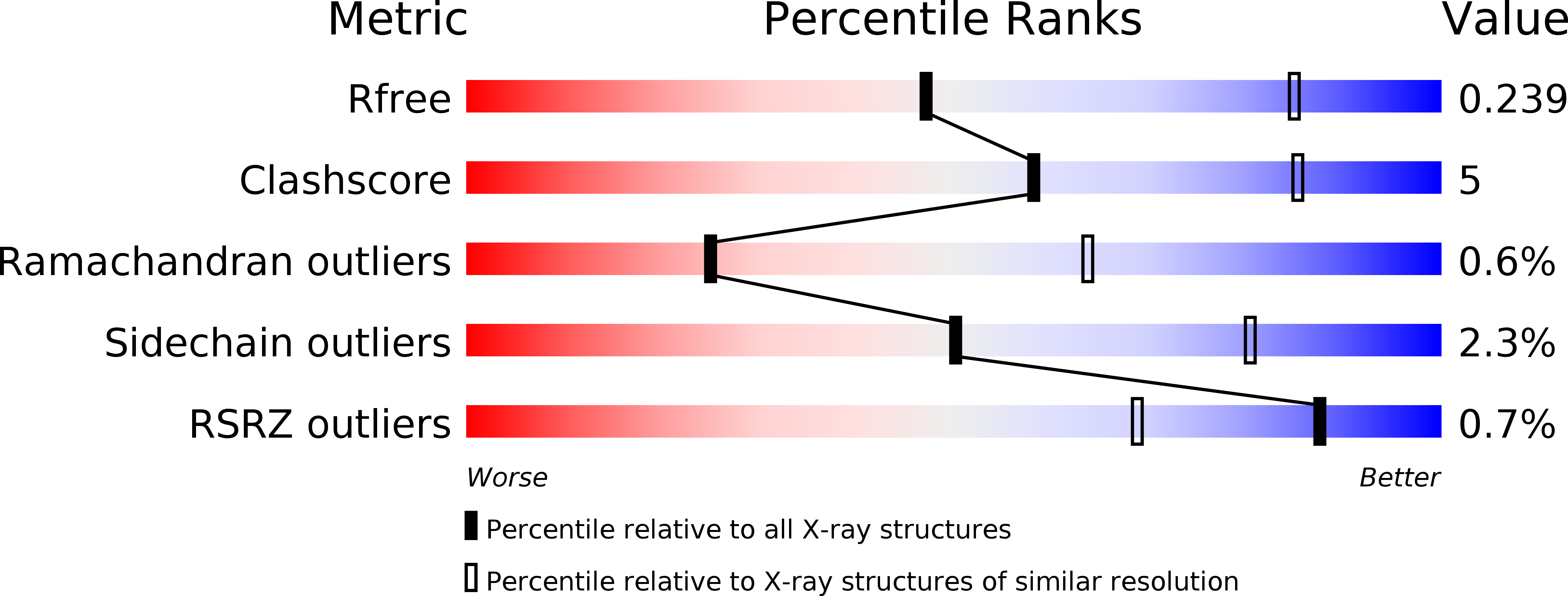
wwPDB Validation   3D Report Full Report


This is version 1.1 of the entry. See complete history


Literature

Structure of the essential peptidoglycan amidotransferase MurT/GatD complex from Streptococcus pneumoniae.

Morlot, C.Straume, D.Peters, K.Hegnar, O.A.Simon, N.Villard, A.M.Contreras-Martel, C.Leisico, F.Breukink, E.Gravier-Pelletier, C.Le Corre, L.Vollmer, W.Pietrancosta, N.Havarstein, L.S.Zapun, A.

(2018) Nat Commun 9: 3180-3180

  • DOI: https://doi.org/10.1038/s41467-018-05602-w
  • Primary Citation of Related Structures:  
    6FQB

  • PubMed Abstract: 

    The universality of peptidoglycan in bacteria underlies the broad spectrum of many successful antibiotics. However, in our times of widespread resistance, the diversity of peptidoglycan modifications offers a variety of new antibacterials targets. In some Gram-positive species such as Streptococcus pneumoniae, Staphylococcus aureus, or Mycobacterium tuberculosis, the second residue of the peptidoglycan precursor, D-glutamate, is amidated into iso-D-glutamine by the essential amidotransferase MurT/GatD complex. Here, we present the structure of this complex at 3.0 Å resolution. MurT has central and C-terminal domains similar to Mur ligases with a cysteine-rich insertion, which probably binds zinc, contributing to the interface with GatD. The mechanism of amidation by MurT is likely similar to the condensation catalyzed by Mur ligases. GatD is a glutaminase providing ammonia that is likely channeled to the MurT active site through a cavity network. The structure and assay presented here constitute a knowledge base for future drug development studies.


  • Organizational Affiliation

    Université Grenoble Alpes, CNRS, CEA, IBS UMR 5075, 38044, Grenoble, France.


Macromolecules
Find similar proteins by:  (by identity cutoff)  |  3D Structure
Entity ID: 1
MoleculeChains Sequence LengthOrganismDetailsImage
Mur ligase family proteinA,
C [auth B],
E [auth C],
G [auth D]
465Streptococcus pneumoniaeMutation(s): 0 
Gene Names: A4260_05845ERS019420_00606ERS020535_00125ERS021368_00525ERS022199_00743ERS043879_01366
EC: 6.3.2
UniProt
Find proteins for Q8DNZ9 (Streptococcus pneumoniae (strain ATCC BAA-255 / R6))
Explore Q8DNZ9 
Go to UniProtKB:  Q8DNZ9
Entity Groups  
Sequence Clusters30% Identity50% Identity70% Identity90% Identity95% Identity100% Identity
UniProt GroupQ8DNZ9
Sequence Annotations
Expand
  • Reference Sequence
Find similar proteins by:  (by identity cutoff)  |  3D Structure
Entity ID: 2
MoleculeChains Sequence LengthOrganismDetailsImage
Cobyric acid synthaseB [auth E],
D [auth F],
F [auth G],
H
260Streptococcus pneumoniaeMutation(s): 0 
Gene Names: 
EC: 6.3.5.10
UniProt
Find proteins for Q8DNZ8 (Streptococcus pneumoniae (strain ATCC BAA-255 / R6))
Explore Q8DNZ8 
Go to UniProtKB:  Q8DNZ8
Entity Groups  
Sequence Clusters30% Identity50% Identity70% Identity90% Identity95% Identity100% Identity
UniProt GroupQ8DNZ8
Sequence Annotations
Expand
  • Reference Sequence
Experimental Data & Validation

Experimental Data

  • Method: X-RAY DIFFRACTION
  • Resolution: 3.00 Å
  • R-Value Free: 0.228 
  • R-Value Work: 0.189 
  • R-Value Observed: 0.193 
  • Space Group: H 3
Unit Cell:
Length ( Å )Angle ( ˚ )
a = 288.43α = 90
b = 288.43β = 90
c = 115.1γ = 120
Software Package:
Software NamePurpose
REFMACrefinement
XDSdata reduction
XSCALEdata scaling
SHARPphasing

Structure Validation

View Full Validation Report


Entry History & Funding Information

Deposition Data


Funding OrganizationLocationGrant Number
INSERM-Astra ZenecaFrance--

Revision History  (Full details and data files)

  • Version 1.0: 2018-08-22
    Type: Initial release
  • Version 1.1: 2018-11-07
    Changes: Data collection