6FNU

Structure of S. cerevisiae Methylenetetrahydrofolate reductase 1, catalytic domain


Experimental Data Snapshot

  • Method: X-RAY DIFFRACTION
  • Resolution: 1.56 Å
  • R-Value Free: 0.190 
  • R-Value Work: 0.152 
  • R-Value Observed: 0.154 

wwPDB Validation   3D Report Full Report


Ligand Structure Quality Assessment 


This is version 1.1 of the entry. See complete history


Literature

Structural basis for the regulation of human 5,10-methylenetetrahydrofolate reductase by phosphorylation and S-adenosylmethionine inhibition.

Froese, D.S.Kopec, J.Rembeza, E.Bezerra, G.A.Oberholzer, A.E.Suormala, T.Lutz, S.Chalk, R.Borkowska, O.Baumgartner, M.R.Yue, W.W.

(2018) Nat Commun 9: 2261-2261

  • DOI: https://doi.org/10.1038/s41467-018-04735-2
  • Primary Citation of Related Structures:  
    6FCX, 6FNU

  • PubMed Abstract: 

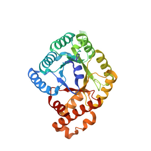
    The folate and methionine cycles are crucial for biosynthesis of lipids, nucleotides and proteins, and production of the methyl donor S-adenosylmethionine (SAM). 5,10-methylenetetrahydrofolate reductase (MTHFR) represents a key regulatory connection between these cycles, generating 5-methyltetrahydrofolate for initiation of the methionine cycle, and undergoing allosteric inhibition by its end product SAM. Our 2.5 Å resolution crystal structure of human MTHFR reveals a unique architecture, appending the well-conserved catalytic TIM-barrel to a eukaryote-only SAM-binding domain. The latter domain of novel fold provides the predominant interface for MTHFR homo-dimerization, positioning the N-terminal serine-rich phosphorylation region near the C-terminal SAM-binding domain. This explains how MTHFR phosphorylation, identified on 11 N-terminal residues (16 in total), increases sensitivity to SAM binding and inhibition. Finally, we demonstrate that the 25-amino-acid inter-domain linker enables conformational plasticity and propose it to be a key mediator of SAM regulation. Together, these results provide insight into the molecular regulation of MTHFR.


  • Organizational Affiliation

    Division of Metabolism and Children's Research Center, University Children's Hospital, CH-8032, Zürich, Switzerland. sean.froese@kispi.uzh.ch.


Macromolecules
Find similar proteins by:  (by identity cutoff)  |  3D Structure
Entity ID: 1
MoleculeChains Sequence LengthOrganismDetailsImage
Methylenetetrahydrofolate reductase 1308Saccharomyces cerevisiaeMutation(s): 0 
Gene Names: MET12YPL023CLPB8C
EC: 1.5.1.20
UniProt
Find proteins for P46151 (Saccharomyces cerevisiae (strain ATCC 204508 / S288c))
Explore P46151 
Go to UniProtKB:  P46151
Entity Groups  
Sequence Clusters30% Identity50% Identity70% Identity90% Identity95% Identity100% Identity
UniProt GroupP46151
Sequence Annotations
Expand
  • Reference Sequence
Small Molecules
Ligands 1 Unique
IDChains Name / Formula / InChI Key2D Diagram3D Interactions
FAD
Query on FAD

Download Ideal Coordinates CCD File 
B [auth A]FLAVIN-ADENINE DINUCLEOTIDE
C27 H33 N9 O15 P2
VWWQXMAJTJZDQX-UYBVJOGSSA-N
Experimental Data & Validation

Experimental Data

  • Method: X-RAY DIFFRACTION
  • Resolution: 1.56 Å
  • R-Value Free: 0.190 
  • R-Value Work: 0.152 
  • R-Value Observed: 0.154 
  • Space Group: P 21 21 2
Unit Cell:
Length ( Å )Angle ( ˚ )
a = 110.65α = 90
b = 54.53β = 90
c = 61.92γ = 90
Software Package:
Software NamePurpose
REFMACrefinement
SCALAdata scaling
PDB_EXTRACTdata extraction
xia2data reduction
PHASERphasing

Structure Validation

View Full Validation Report


Ligand Structure Quality Assessment 


Entry History 

Revision History  (Full details and data files)

  • Version 1.0: 2018-03-07
    Type: Initial release
  • Version 1.1: 2018-07-04
    Changes: Data collection, Database references