Cep120 promotes microtubule formation through a unique tubulin binding C2 domain.
Sharma, A., Gerard, S.F., Olieric, N., Steinmetz, M.O.(2018) J Struct Biol 203: 62-70
- PubMed: 29398280
- DOI: https://doi.org/10.1016/j.jsb.2018.01.009
- Primary Citation of Related Structures:
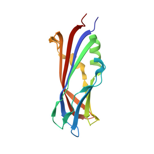
6FLJ, 6FLK - PubMed Abstract:
Centrioles are microtubule-based structures that play essential roles in cell division and cilia biogenesis. Cep120 is an important protein for correct centriole formation and mutations in the Cep120 gene cause severe human diseases like Joubert syndrome and complex ciliopathies. Here, we show that Cep120 contains three consecutive C2 domains that are followed by a coiled-coil dimerization domain. Surprisingly, unlike the classical C2 domains, all three Cep120 C2 domains lack calcium- and phospholipid-binding activities. However, biophysical and biochemical assays revealed that the N-terminal Cep120 C2 domain (C2A) binds to both tubulin and microtubules, and promotes microtubule formation. Structural analyses coupled with mutagenesis identified a highly conserved, positively charged residue patch on the surface of Cep120 C2A, which mediates the interaction with tubulin and microtubules. Together, our results establish Cep120 C2A as a unique microtubule-binding domain. They further provide insights into the molecular mechanism of Cep120 during centriole biogenesis.
Organizational Affiliation:
Laboratory of Biomolecular Research, Division of Biology and Chemistry, Paul Scherrer Institut, 5232 Villigen PSI, Switzerland.