6FKN

Drosophila Plexin A in complex with Semaphorin 1b


Experimental Data Snapshot

  • Method: X-RAY DIFFRACTION
  • Resolution: 4.80 Å
  • R-Value Free: 0.297 
  • R-Value Work: 0.285 
  • R-Value Observed: 0.285 

wwPDB Validation   3D Report Full Report


This is version 2.1 of the entry. See complete history


Literature

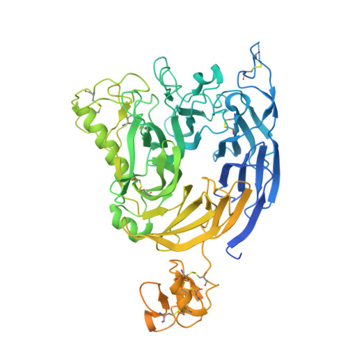
Structural basis of semaphorin-plexin cis interaction.

Rozbesky, D.Verhagen, M.G.Karia, D.Nagy, G.N.Alvarez, L.Robinson, R.A.Harlos, K.Padilla-Parra, S.Pasterkamp, R.J.Jones, E.Y.

(2020) EMBO J : e102926-e102926

  • DOI: https://doi.org/10.15252/embj.2019102926
  • Primary Citation of Related Structures:  
    6FKM, 6FKN

  • PubMed Abstract: 

    Semaphorin ligands interact with plexin receptors to contribute to functions in the development of myriad tissues including neurite guidance and synaptic organisation within the nervous system. Cell-attached semaphorins interact in trans with plexins on opposing cells, but also in cis on the same cell. The interplay between trans and cis interactions is crucial for the regulated development of complex neural circuitry, but the underlying molecular mechanisms are uncharacterised. We have discovered a distinct mode of interaction through which the Drosophila semaphorin Sema1b and mouse Sema6A mediate binding in cis to their cognate plexin receptors. Our high-resolution structural, biophysical and in vitro analyses demonstrate that monomeric semaphorins can mediate a distinctive plexin binding mode. These findings suggest the interplay between monomeric vs dimeric states has a hereto unappreciated role in semaphorin biology, providing a mechanism by which Sema6s may balance cis and trans functionalities.


  • Organizational Affiliation

    Division of Structural Biology, Wellcome Centre for Human Genetics, University of Oxford, Oxford, UK.


Macromolecules
Find similar proteins by:  (by identity cutoff)  |  3D Structure
Entity ID: 1
MoleculeChains Sequence LengthOrganismDetailsImage
Plexin A, isoform A
A, C
715Drosophila melanogasterMutation(s): 0 
Gene Names: 
UniProt
Find proteins for Q9V491 (Drosophila melanogaster)
Explore Q9V491 
Go to UniProtKB:  Q9V491
Entity Groups  
Sequence Clusters30% Identity50% Identity70% Identity90% Identity95% Identity100% Identity
UniProt GroupQ9V491
Sequence Annotations
Expand
  • Reference Sequence
Find similar proteins by:  (by identity cutoff)  |  3D Structure
Entity ID: 2
MoleculeChains Sequence LengthOrganismDetailsImage
MIP07328p
B, D
578Drosophila melanogasterMutation(s): 0 
Gene Names: Sema1bSema-1bSema-1b-RBsemaphorin-likeCG6446Dmel_CG6446
UniProt
Find proteins for Q7KK54 (Drosophila melanogaster)
Explore Q7KK54 
Go to UniProtKB:  Q7KK54
Entity Groups  
Sequence Clusters30% Identity50% Identity70% Identity90% Identity95% Identity100% Identity
UniProt GroupQ7KK54
Sequence Annotations
Expand
  • Reference Sequence
Oligosaccharides

Help

Entity ID: 3
MoleculeChains Length2D Diagram Glycosylation3D Interactions
alpha-D-mannopyranose-(1-3)-beta-D-mannopyranose-(1-4)-2-acetamido-2-deoxy-beta-D-glucopyranose-(1-4)-2-acetamido-2-deoxy-beta-D-glucopyranose
E
4N-Glycosylation
Glycosylation Resources
GlyTouCan:  G81315DD
GlyCosmos:  G81315DD
GlyGen:  G81315DD
Entity ID: 4
MoleculeChains Length2D Diagram Glycosylation3D Interactions
beta-D-mannopyranose-(1-4)-2-acetamido-2-deoxy-beta-D-glucopyranose-(1-4)-2-acetamido-2-deoxy-beta-D-glucopyranose
F
3N-Glycosylation
Glycosylation Resources
GlyTouCan:  G15407YE
GlyCosmos:  G15407YE
GlyGen:  G15407YE
Entity ID: 5
MoleculeChains Length2D Diagram Glycosylation3D Interactions
alpha-D-mannopyranose-(1-3)-[alpha-D-mannopyranose-(1-6)]beta-D-mannopyranose-(1-4)-2-acetamido-2-deoxy-beta-D-glucopyranose-(1-4)-2-acetamido-2-deoxy-beta-D-glucopyranose
G, H
5N-Glycosylation
Glycosylation Resources
GlyTouCan:  G22768VO
GlyCosmos:  G22768VO
GlyGen:  G22768VO
Experimental Data & Validation

Experimental Data

  • Method: X-RAY DIFFRACTION
  • Resolution: 4.80 Å
  • R-Value Free: 0.297 
  • R-Value Work: 0.285 
  • R-Value Observed: 0.285 
  • Space Group: P 65 2 2
Unit Cell:
Length ( Å )Angle ( ˚ )
a = 153.63α = 90
b = 153.63β = 90
c = 425.37γ = 120
Software Package:
Software NamePurpose
PHENIXrefinement
xia2data reduction
xia2data scaling
PHASERphasing

Structure Validation

View Full Validation Report


Entry History & Funding Information

Deposition Data


Funding OrganizationLocationGrant Number
Cancer Research UKUnited KingdomB5R00120

Revision History  (Full details and data files)

  • Version 1.0: 2019-02-06
    Type: Initial release
  • Version 1.1: 2020-06-17
    Changes: Data collection, Database references
  • Version 2.0: 2020-07-29
    Type: Remediation
    Reason: Carbohydrate remediation
    Changes: Advisory, Atomic model, Data collection, Derived calculations, Structure summary
  • Version 2.1: 2024-01-17
    Changes: Data collection, Database references, Derived calculations, Refinement description, Structure summary