6FHT

Crystal structure of an artificial phytochrome regulated adenylate/guanylate cyclase in its dark adapted Pr form


Experimental Data Snapshot

  • Method: X-RAY DIFFRACTION
  • Resolution: 2.35 Å
  • R-Value Free: 0.227 
  • R-Value Work: 0.180 
  • R-Value Observed: 0.182 

wwPDB Validation   3D Report Full Report


Ligand Structure Quality Assessment 


This is version 1.3 of the entry. See complete history


Literature

Structure-guided design and functional characterization of an artificial red light-regulated guanylate/adenylate cyclase for optogenetic applications.

Etzl, S.Lindner, R.Nelson, M.D.Winkler, A.

(2018) J Biol Chem 293: 9078-9089

  • DOI: https://doi.org/10.1074/jbc.RA118.003069
  • Primary Citation of Related Structures:  
    6FHT

  • PubMed Abstract: 

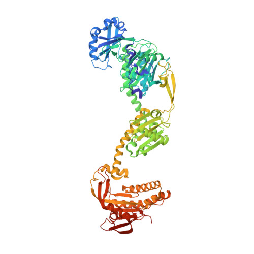
    Genetically targeting biological systems to control cellular processes with light is the concept of optogenetics. Despite impressive developments in this field, underlying molecular mechanisms of signal transduction of the employed photoreceptor modules are frequently not sufficiently understood to rationally design new optogenetic tools. Here, we investigate the requirements for functional coupling of red light-sensing phytochromes with non-natural enzymatic effectors by creating a series of constructs featuring the Deinococcus radiodurans bacteriophytochrome linked to a Synechocystis guanylate/adenylate cyclase. Incorporating characteristic structural elements important for cyclase regulation in our designs, we identified several red light-regulated fusions with promising properties. We provide details of one light-activated construct with low dark-state activity and high dynamic range that outperforms previous optogenetic tools in vitro and expands our in vivo toolkit, as demonstrated by manipulation of Caenorhabditis elegans locomotor activity. The full-length crystal structure of this phytochrome-linked cyclase revealed molecular details of photoreceptor-effector coupling, highlighting the importance of the regulatory cyclase element. Analysis of conformational dynamics by hydrogen-deuterium exchange in different functional states enriched our understanding of phytochrome signaling and signal integration by effectors. We found that light-induced conformational changes in the phytochrome destabilize the coiled-coil sensor-effector linker, which releases the cyclase regulatory element from an inhibited conformation, increasing cyclase activity of this artificial system. Future designs of optogenetic functionalities may benefit from our work, indicating that rational considerations for the effector improve the rate of success of initial designs to obtain optogenetic tools with superior properties.


  • Organizational Affiliation

    From the Institute of Biochemistry, Graz University of Technology, Petersgasse 12/II, 8010 Graz, Austria.


Macromolecules
Find similar proteins by:  (by identity cutoff)  |  3D Structure
Entity ID: 1
MoleculeChains Sequence LengthOrganismDetailsImage
Bacteriophytochrome,Adenylate cyclase
A, B
741Deinococcus radioduransSynechocystis sp. PCC 6803 substr. Kazusa
This entity is chimeric
Mutation(s): 1 
Gene Names: bphPDR_A0050cyaA
EC: 2.7.13.3
UniProt
Find proteins for Q9RZA4 (Deinococcus radiodurans (strain ATCC 13939 / DSM 20539 / JCM 16871 / CCUG 27074 / LMG 4051 / NBRC 15346 / NCIMB 9279 / VKM B-1422 / R1))
Explore Q9RZA4 
Go to UniProtKB:  Q9RZA4
Find proteins for P72951 (Synechocystis sp. (strain PCC 6803 / Kazusa))
Explore P72951 
Go to UniProtKB:  P72951
Entity Groups  
Sequence Clusters30% Identity50% Identity70% Identity90% Identity95% Identity100% Identity
UniProt GroupsQ9RZA4P72951
Sequence Annotations
Expand
  • Reference Sequence
Small Molecules
Ligands 3 Unique
IDChains Name / Formula / InChI Key2D Diagram3D Interactions
LBV
Query on LBV

Download Ideal Coordinates CCD File 
C [auth A],
K [auth B]
3-[2-[(Z)-[3-(2-carboxyethyl)-5-[(Z)-(4-ethenyl-3-methyl-5-oxidanylidene-pyrrol-2-ylidene)methyl]-4-methyl-pyrrol-1-ium -2-ylidene]methyl]-5-[(Z)-[(3E)-3-ethylidene-4-methyl-5-oxidanylidene-pyrrolidin-2-ylidene]methyl]-4-methyl-1H-pyrrol-3- yl]propanoic acid
C33 H37 N4 O6
DKMLMZVDTGOEGU-ISEYCTJISA-O
12P
Query on 12P

Download Ideal Coordinates CCD File 
J [auth A]DODECAETHYLENE GLYCOL
C24 H50 O13
WRZXKWFJEFFURH-UHFFFAOYSA-N
SO4
Query on SO4

Download Ideal Coordinates CCD File 
D [auth A]
E [auth A]
F [auth A]
G [auth A]
H [auth A]
D [auth A],
E [auth A],
F [auth A],
G [auth A],
H [auth A],
I [auth A],
L [auth B],
M [auth B],
N [auth B],
O [auth B]
SULFATE ION
O4 S
QAOWNCQODCNURD-UHFFFAOYSA-L
Experimental Data & Validation

Experimental Data

  • Method: X-RAY DIFFRACTION
  • Resolution: 2.35 Å
  • R-Value Free: 0.227 
  • R-Value Work: 0.180 
  • R-Value Observed: 0.182 
  • Space Group: C 1 2 1
Unit Cell:
Length ( Å )Angle ( ˚ )
a = 286.19α = 90
b = 108.82β = 100.84
c = 67.99γ = 90
Software Package:
Software NamePurpose
XDSdata reduction
XSCALEdata scaling
PHASERphasing
PHENIXrefinement
PDB_EXTRACTdata extraction

Structure Validation

View Full Validation Report


Ligand Structure Quality Assessment 


Entry History & Funding Information

Deposition Data


Funding OrganizationLocationGrant Number
Austrian Science FundAustriaP27124

Revision History  (Full details and data files)

  • Version 1.0: 2018-04-18
    Type: Initial release
  • Version 1.1: 2018-05-09
    Changes: Data collection, Database references, Source and taxonomy
  • Version 1.2: 2018-06-20
    Changes: Data collection, Database references, Derived calculations
  • Version 1.3: 2024-01-17
    Changes: Data collection, Database references, Derived calculations, Refinement description, Structure summary