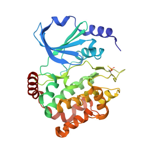
6FD3

Thiophosphorylated PAK3 kinase domain


Experimental Data Snapshot

  • Method: X-RAY DIFFRACTION
  • Resolution: 1.52 Å
  • R-Value Free: 0.190 
  • R-Value Work: 0.175 
  • R-Value Observed: 0.176 

wwPDB Validation   3D Report Full Report


Ligand Structure Quality Assessment 


This is version 1.2 of the entry. See complete history


Literature

Solution structures and biophysical analysis of full-length group A PAKs reveal they are monomeric and auto-inhibited incis.

Sorrell, F.J.Kilian, L.M.Elkins, J.M.

(2019) Biochem J 476: 1037-1051

  • DOI: https://doi.org/10.1042/BCJ20180867
  • Primary Citation of Related Structures:  
    6FD3

  • PubMed Abstract: 

    The group A p21-activated kinases (PAKs) exist in an auto-inhibited form until activated by GTPase binding and auto-phosphorylation. In the auto-inhibited form, a regulatory domain binds to the kinase domain (KD) blocking the binding of substrates, and CDC42 or Rac binding to the regulatory domain relieves this auto-inhibition allowing auto-phosphorylation on the KD activation loop. We have determined the crystal structure of the PAK3 catalytic domain and by small angle X-ray scattering, the solution-phase structures of full-length inactive PAK1 and PAK3. The structures reveal a compact but elongated molecular shape that demonstrates that, together with multiple independent biophysical measurements and in contrast with previous assumptions, group A PAKs are monomeric both before and after activation, consistent with an activation mechanism of cis -auto-inhibition and initial cis -auto-phosphorylation, followed by transient dimerisation to allow trans -auto-phosphorylation for full activation, yielding a monomeric active PAK protein.


  • Organizational Affiliation

    Structural Genomics Consortium, University of Oxford, Old Road Campus Research Building, Roosevelt Drive, Oxford OX3 7DQ, U.K.


Macromolecules
Find similar proteins by:  (by identity cutoff)  |  3D Structure
Entity ID: 1
MoleculeChains Sequence LengthOrganismDetailsImage
Serine/threonine-protein kinase PAK 3300Homo sapiensMutation(s): 1 
Gene Names: PAK3OPHN3
EC: 2.7.11.1
UniProt & NIH Common Fund Data Resources
Find proteins for O75914 (Homo sapiens)
Explore O75914 
Go to UniProtKB:  O75914
PHAROS:  O75914
GTEx:  ENSG00000077264 
Entity Groups  
Sequence Clusters30% Identity50% Identity70% Identity90% Identity95% Identity100% Identity
UniProt GroupO75914
Sequence Annotations
Expand
  • Reference Sequence
Experimental Data & Validation

Experimental Data

  • Method: X-RAY DIFFRACTION
  • Resolution: 1.52 Å
  • R-Value Free: 0.190 
  • R-Value Work: 0.175 
  • R-Value Observed: 0.176 
  • Space Group: I 41
Unit Cell:
Length ( Å )Angle ( ˚ )
a = 108.825α = 90
b = 108.825β = 90
c = 57.934γ = 90
Software Package:
Software NamePurpose
PHENIXrefinement
XDSdata reduction
Aimlessdata scaling
PHASERphasing
PDB_EXTRACTdata extraction

Structure Validation

View Full Validation Report


Ligand Structure Quality Assessment 


Entry History 

Deposition Data

Revision History  (Full details and data files)

  • Version 1.0: 2018-01-03
    Type: Initial release
  • Version 1.1: 2019-07-17
    Changes: Data collection, Database references
  • Version 1.2: 2024-01-17
    Changes: Data collection, Database references, Derived calculations, Refinement description