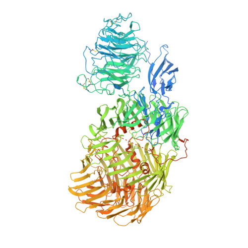
6FAY

Teneurin3 monomer


Experimental Data Snapshot

  • Method: ELECTRON MICROSCOPY
  • Resolution: 3.80 Å
  • Aggregation State: PARTICLE 
  • Reconstruction Method: SINGLE PARTICLE 

wwPDB Validation   3D Report Full Report


This is version 1.2 of the entry. See complete history


Literature

Structures of Teneurin adhesion receptors reveal an ancient fold for cell-cell interaction.

Jackson, V.A.Meijer, D.H.Carrasquero, M.van Bezouwen, L.S.Lowe, E.D.Kleanthous, C.Janssen, B.J.C.Seiradake, E.

(2018) Nat Commun 9: 1079-1079

  • DOI: https://doi.org/10.1038/s41467-018-03460-0
  • Primary Citation of Related Structures:  
    6FAY, 6FB3

  • PubMed Abstract: 

    Teneurins are ancient cell-cell adhesion receptors that are vital for brain development and synapse organisation. They originated in early metazoan evolution through a horizontal gene transfer event when a bacterial YD-repeat toxin fused to a eukaryotic receptor. We present X-ray crystallography and cryo-EM structures of two Teneurins, revealing a ~200 kDa extracellular super-fold in which eight sub-domains form an intricate structure centred on a spiralling YD-repeat shell. An alternatively spliced loop, which is implicated in homophilic Teneurin interaction and specificity, is exposed and thus poised for interaction. The N-terminal side of the shell is 'plugged' via a fibronectin-plug domain combination, which defines a new class of YD proteins. Unexpectedly, we find that these proteins are widespread amongst modern bacteria, suggesting early metazoan receptor evolution from a distinct class of proteins, which today includes both bacterial proteins and eukaryotic Teneurins.


  • Organizational Affiliation

    Department of Biochemistry, Oxford University, OX1 3QU, Oxford, UK. verity.jackson@magd.ox.ac.uk.


Macromolecules
Find similar proteins by:  (by identity cutoff)  |  3D Structure
Entity ID: 1
MoleculeChains Sequence LengthOrganismDetailsImage
Odz3 protein1,876Mus musculusMutation(s): 0 
Gene Names: Tenm3Odz3
UniProt
Find proteins for Q9WTS6 (Mus musculus)
Explore Q9WTS6 
Go to UniProtKB:  Q9WTS6
Entity Groups  
Sequence Clusters30% Identity50% Identity70% Identity90% Identity95% Identity100% Identity
UniProt GroupQ9WTS6
Sequence Annotations
Expand
  • Reference Sequence
Experimental Data & Validation

Experimental Data

  • Method: ELECTRON MICROSCOPY
  • Resolution: 3.80 Å
  • Aggregation State: PARTICLE 
  • Reconstruction Method: SINGLE PARTICLE 
EM Software:
TaskSoftware PackageVersion
RECONSTRUCTIONcryoSPARC
MODEL REFINEMENTPHENIX

Structure Validation

View Full Validation Report


Entry History & Funding Information

Deposition Data


Funding OrganizationLocationGrant Number
European Molecular Biology OrganizationNetherlands125-2015
Netherlands Organisation for Scientific ResearchNetherlands723.012.002

Revision History  (Full details and data files)

  • Version 1.0: 2018-03-28
    Type: Initial release
  • Version 1.1: 2019-12-11
    Changes: Other
  • Version 1.2: 2020-07-29
    Type: Remediation
    Reason: Carbohydrate remediation
    Changes: Data collection, Derived calculations, Structure summary