6F7T

Crystal Structure of an Fab fragment in complex with a peptide from Bacillus subtilis RNase Y


Experimental Data Snapshot

  • Method: X-RAY DIFFRACTION
  • Resolution: 2.60 Å
  • R-Value Free: 0.253 
  • R-Value Work: 0.202 
  • R-Value Observed: 0.204 

wwPDB Validation   3D Report Full Report


This is version 1.3 of the entry. See complete history


Literature

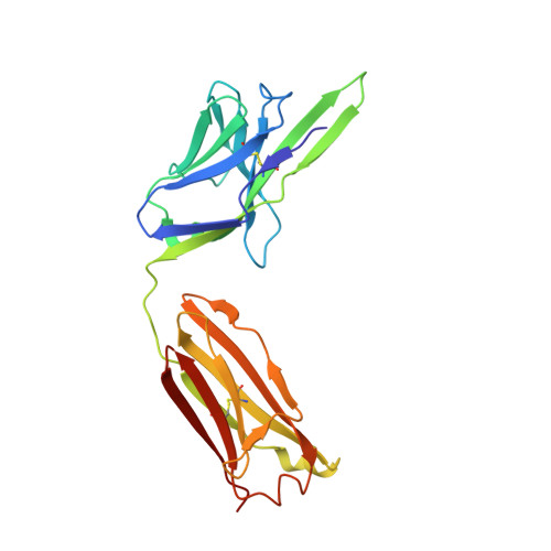
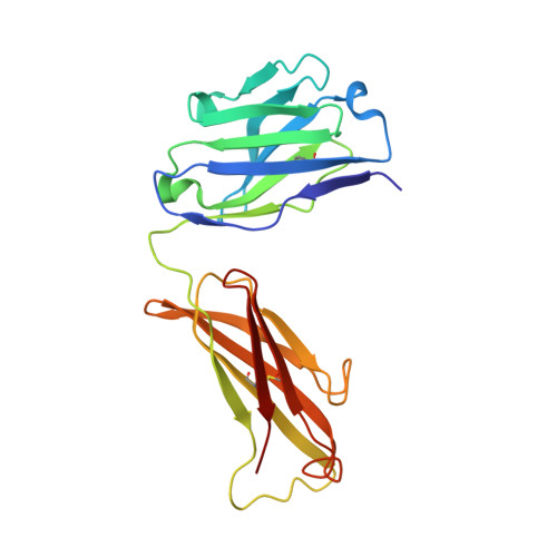
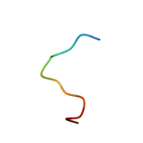
Dissociation of the Dimer of the Intrinsically Disordered Domain of RNase Y upon Antibody Binding.

Hardouin, P.Velours, C.Bou-Nader, C.Assrir, N.Laalami, S.Putzer, H.Durand, D.Golinelli-Pimpaneau, B.

(2018) Biophys J 115: 2102-2113

  • DOI: https://doi.org/10.1016/j.bpj.2018.10.016
  • Primary Citation of Related Structures:  
    6F7T

  • PubMed Abstract: 

    Although RNase Y acts as the key enzyme initiating messenger RNA decay in Bacillus subtilis and likely in many other Gram-positive bacteria, its three-dimensional structure remains unknown. An antibody belonging to the rare immunoglobulin G (IgG) 2b λx isotype was raised against a 12-residue conserved peptide from the N-terminal noncatalytic domain of B. subtilis RNase Y (BsRNaseY) that is predicted to be intrinsically disordered. Here, we show that this domain can be produced as a stand-alone protein called Nter-BsRNaseY that undergoes conformational changes between monomeric and dimeric forms. Circular dichroism and size exclusion chromatography coupled with multiangle light scattering or with small angle x-ray scattering indicate that the Nter-BsRNaseY dimer displays an elongated form and a high content of α-helices, in agreement with the existence of a central coiled-coil structure appended with flexible ends, and that the monomeric state of Nter-BsRNaseY is favored upon binding the fragment antigen binding (Fab) of the antibody. The dissociation constants of the IgG/BsRNaseY, IgG/Nter-BsRNaseY, and IgG/peptide complexes indicate that the affinity of the IgG for Nter-BsRNaseY is in the nM range and suggest that the peptide is less accessible in BsRNaseY than in Nter-BsRNaseY. The crystal structure of the Fab in complex with the peptide antigen shows that the peptide adopts an elongated U-shaped conformation in which the unique hydrophobic residue of the peptide, Leu6, is completely buried. The peptide/Fab complex may mimic the interaction of a microdomain of the N-terminal domain of BsRNaseY with one of its cellular partners within the degradosome complex. Altogether, our results suggest that BsRNaseY may become accessible for protein interaction upon dissociation of its N-terminal domain into the monomeric form.


  • Organizational Affiliation

    Laboratoire de Chimie des Processus Biologiques, UMR 8229 CNRS, Collège de France, Université Pierre et Marie Curie, Paris CEDEX 05, France.


Macromolecules
Find similar proteins by:  (by identity cutoff)  |  3D Structure
Entity ID: 1
MoleculeChains Sequence LengthOrganismDetailsImage
FAB RY79-90, LIGHT CHAINA,
F [auth L]
218Mus musculusMutation(s): 0 
Entity Groups  
Sequence Clusters30% Identity50% Identity70% Identity90% Identity95% Identity100% Identity
Sequence Annotations
Expand
  • Reference Sequence
Find similar proteins by:  (by identity cutoff)  |  3D Structure
Entity ID: 2
MoleculeChains Sequence LengthOrganismDetailsImage
FAB RY79-90, HEAVY CHAINB,
E [auth H]
211Mus musculusMutation(s): 0 
Entity Groups  
Sequence Clusters30% Identity50% Identity70% Identity90% Identity95% Identity100% Identity
Sequence Annotations
Expand
  • Reference Sequence

Find similar proteins by:  Sequence   |   3D Structure  

Entity ID: 3
MoleculeChains Sequence LengthOrganismDetailsImage
Ribonuclease Y
C, D
12Bacillus subtilisMutation(s): 0 
EC: 3.1
UniProt
Find proteins for O31774 (Bacillus subtilis (strain 168))
Explore O31774 
Go to UniProtKB:  O31774
Entity Groups  
Sequence Clusters30% Identity50% Identity70% Identity90% Identity95% Identity100% Identity
UniProt GroupO31774
Sequence Annotations
Expand
  • Reference Sequence
Small Molecules
Ligands 1 Unique
IDChains Name / Formula / InChI Key2D Diagram3D Interactions
MG
Query on MG

Download Ideal Coordinates CCD File 
G [auth L]MAGNESIUM ION
Mg
JLVVSXFLKOJNIY-UHFFFAOYSA-N
Experimental Data & Validation

Experimental Data

  • Method: X-RAY DIFFRACTION
  • Resolution: 2.60 Å
  • R-Value Free: 0.253 
  • R-Value Work: 0.202 
  • R-Value Observed: 0.204 
  • Space Group: P 21 21 2
Unit Cell:
Length ( Å )Angle ( ˚ )
a = 98.68α = 90
b = 130.45β = 90
c = 72.05γ = 90
Software Package:
Software NamePurpose
BUSTERrefinement
XDSdata reduction
XSCALEdata scaling
PHASERphasing

Structure Validation

View Full Validation Report


Entry History & Funding Information

Deposition Data


Funding OrganizationLocationGrant Number
LABEX DynamoFranceANR-11-LABX-0011
French National Research AgencyFranceANR-11-BSV8-026

Revision History  (Full details and data files)

  • Version 1.0: 2018-10-31
    Type: Initial release
  • Version 1.1: 2018-11-28
    Changes: Data collection, Database references
  • Version 1.2: 2018-12-19
    Changes: Data collection, Database references
  • Version 1.3: 2024-01-17
    Changes: Data collection, Database references, Refinement description