6F5M

Crystal structure of highly glycosylated human leukocyte elastase in complex with a thiazolidinedione inhibitor


Experimental Data Snapshot

  • Method: X-RAY DIFFRACTION
  • Resolution: 2.70 Å
  • R-Value Free: 0.234 
  • R-Value Work: 0.176 
  • R-Value Observed: 0.180 

wwPDB Validation   3D Report Full Report


Ligand Structure Quality Assessment 


This is version 2.1 of the entry. See complete history


Literature

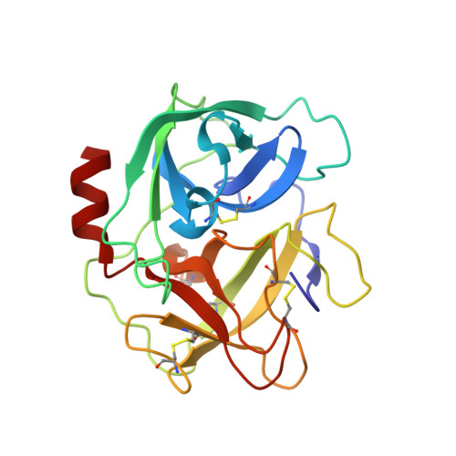
Crystal structure of highly glycosylated human leukocyte elastase in complex with an S2' site binding inhibitor.

Hochscherf, J.Pietsch, M.Tieu, W.Kuan, K.Abell, A.D.Gutschow, M.Niefind, K.

(2018) Acta Crystallogr F Struct Biol Commun 74: 480-489

  • DOI: https://doi.org/10.1107/S2053230X1800537X
  • Primary Citation of Related Structures:  
    6F5M

  • PubMed Abstract: 

    Glycosylated human leukocyte elastase (HLE) was crystallized and structurally analysed in complex with a 1,3-thiazolidine-2,4-dione derivative that had been identified as an HLE inhibitor in preliminary studies. In contrast to previously described HLE structures with small-molecule inhibitors, in this structure the inhibitor does not bind to the S1 and S2 substrate-recognition sites; rather, this is the first HLE structure with a synthetic inhibitor in which the S2' site is blocked that normally binds the second side chain at the C-terminal side of the scissile peptide bond in a substrate protein. The inhibitor also induces the formation of crystalline HLE dimers that block access to the active sites and that are also predicted to be stable in solution. Neither such HLE dimers nor the corresponding crystal packing have been observed in previous HLE crystal structures. This novel crystalline environment contributes to the observation that comparatively large parts of the N-glycan chains of HLE are defined by electron density. The final HLE structure contains the largest structurally defined carbohydrate trees among currently available HLE structures.


  • Organizational Affiliation

    Department of Chemistry, Institute of Biochemistry, Universität zu Köln, Zülpicher Str. 47, 50674 Cologne, Germany.


Macromolecules
Find similar proteins by:  (by identity cutoff)  |  3D Structure
Entity ID: 1
MoleculeChains Sequence LengthOrganismDetailsImage
Neutrophil elastase
A, B
218Homo sapiensMutation(s): 0 
EC: 3.4.21.37
UniProt & NIH Common Fund Data Resources
Find proteins for P08246 (Homo sapiens)
Explore P08246 
Go to UniProtKB:  P08246
PHAROS:  P08246
GTEx:  ENSG00000197561 
Entity Groups  
Sequence Clusters30% Identity50% Identity70% Identity90% Identity95% Identity100% Identity
UniProt GroupP08246
Sequence Annotations
Expand
  • Reference Sequence
Oligosaccharides

Help

Entity ID: 2
MoleculeChains Length2D Diagram Glycosylation3D Interactions
alpha-D-mannopyranose-(1-6)-beta-D-mannopyranose-(1-4)-2-acetamido-2-deoxy-beta-D-glucopyranose-(1-4)-[alpha-L-fucopyranose-(1-6)]2-acetamido-2-deoxy-beta-D-glucopyranose
C
5N-Glycosylation
Glycosylation Resources
GlyTouCan:  G00395TQ
GlyCosmos:  G00395TQ
GlyGen:  G00395TQ
Entity ID: 3
MoleculeChains Length2D Diagram Glycosylation3D Interactions
beta-D-mannopyranose-(1-4)-2-acetamido-2-deoxy-beta-D-glucopyranose-(1-4)-[alpha-L-fucopyranose-(1-6)]2-acetamido-2-deoxy-beta-D-glucopyranose
D, F
4N-Glycosylation
Glycosylation Resources
GlyTouCan:  G32152BH
GlyCosmos:  G32152BH
GlyGen:  G32152BH
Entity ID: 4
MoleculeChains Length2D Diagram Glycosylation3D Interactions
alpha-D-mannopyranose-(1-3)-[alpha-D-mannopyranose-(1-6)]beta-D-mannopyranose-(1-4)-2-acetamido-2-deoxy-beta-D-glucopyranose-(1-4)-[alpha-L-fucopyranose-(1-6)]2-acetamido-2-deoxy-beta-D-glucopyranose
E
6N-Glycosylation
Glycosylation Resources
GlyTouCan:  G82348BZ
GlyCosmos:  G82348BZ
GlyGen:  G82348BZ
Small Molecules
Ligands 3 Unique
IDChains Name / Formula / InChI Key2D Diagram3D Interactions
CQH
Query on CQH

Download Ideal Coordinates CCD File 
K [auth A],
L [auth B]
5-[[4-[[(2~{S})-4-methyl-1-oxidanylidene-1-[(2-propylphenyl)amino]pentan-2-yl]carbamoyl]phenyl]methyl]-2-oxidanylidene-1,3-thiazol-1-ium-4-olate
C26 H29 N3 O4 S
NWLPQCZZJXMUOM-NRFANRHFSA-N
SO4
Query on SO4

Download Ideal Coordinates CCD File 
G [auth A],
H [auth A],
M [auth B],
N [auth B]
SULFATE ION
O4 S
QAOWNCQODCNURD-UHFFFAOYSA-L
ACT
Query on ACT

Download Ideal Coordinates CCD File 
I [auth A],
J [auth A],
O [auth B]
ACETATE ION
C2 H3 O2
QTBSBXVTEAMEQO-UHFFFAOYSA-M
Experimental Data & Validation

Experimental Data

  • Method: X-RAY DIFFRACTION
  • Resolution: 2.70 Å
  • R-Value Free: 0.234 
  • R-Value Work: 0.176 
  • R-Value Observed: 0.180 
  • Space Group: H 3 2
Unit Cell:
Length ( Å )Angle ( ˚ )
a = 204.557α = 90
b = 204.557β = 90
c = 62.155γ = 120
Software Package:
Software NamePurpose
PHENIXrefinement
XDSdata reduction
Aimlessdata scaling
PHASERphasing

Structure Validation

View Full Validation Report


Ligand Structure Quality Assessment 


Entry History & Funding Information

Deposition Data


Funding OrganizationLocationGrant Number
German Research FoundationGermanyNI 643/4-1

Revision History  (Full details and data files)

  • Version 1.0: 2018-08-08
    Type: Initial release
  • Version 1.1: 2018-08-15
    Changes: Data collection, Database references
  • Version 2.0: 2020-07-29
    Type: Remediation
    Reason: Carbohydrate remediation
    Changes: Atomic model, Data collection, Derived calculations, Structure summary
  • Version 2.1: 2024-01-17
    Changes: Data collection, Database references, Derived calculations, Refinement description, Structure summary