6F5D

Trypanosoma brucei F1-ATPase


Experimental Data Snapshot

  • Method: X-RAY DIFFRACTION
  • Resolution: 3.20 Å
  • R-Value Free: 0.297 
  • R-Value Work: 0.272 
  • R-Value Observed: 0.273 

wwPDB Validation   3D Report Full Report


Ligand Structure Quality Assessment 


This is version 2.1 of the entry. See complete history


Literature

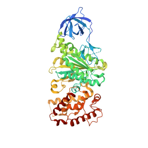
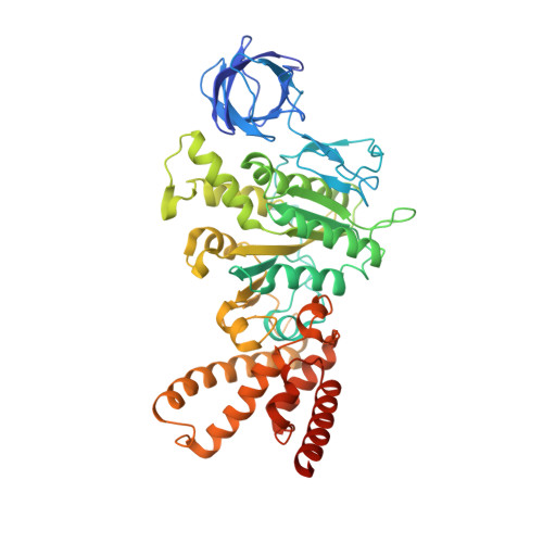
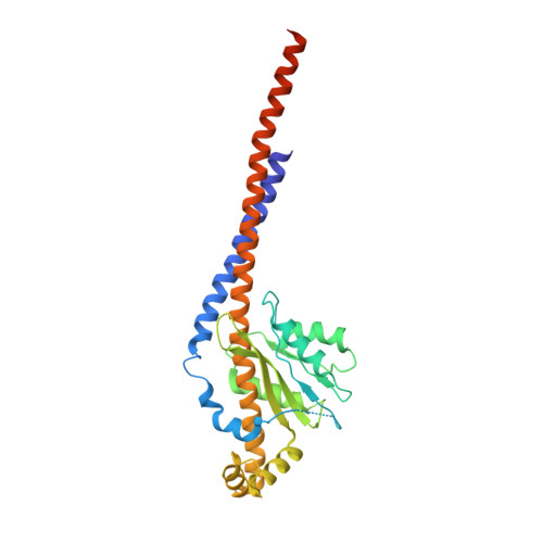
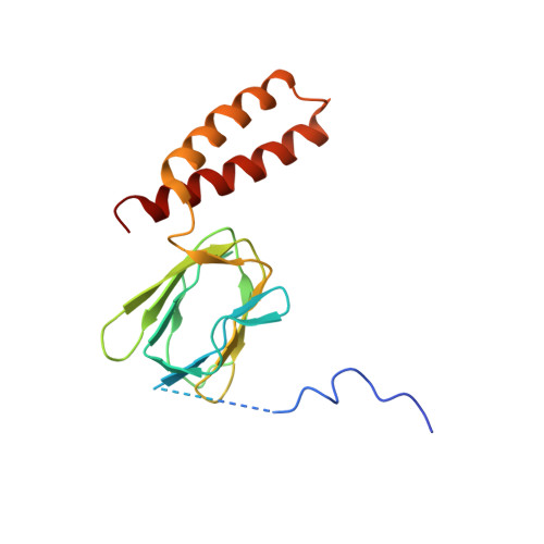
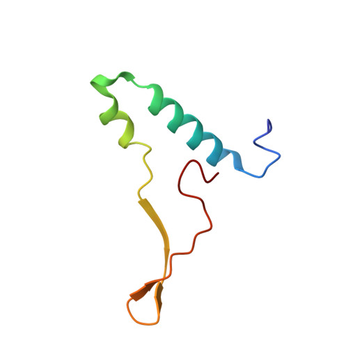
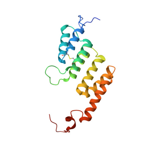
ATP synthase fromTrypanosoma bruceihas an elaborated canonical F1-domain and conventional catalytic sites.

Montgomery, M.G.Gahura, O.Leslie, A.G.W.Zikova, A.Walker, J.E.

(2018) Proc Natl Acad Sci U S A 115: 2102-2107

  • DOI: https://doi.org/10.1073/pnas.1720940115
  • Primary Citation of Related Structures:  
    6F5D

  • PubMed Abstract: 

    The structures and functions of the components of ATP synthases, especially those subunits involved directly in the catalytic formation of ATP, are widely conserved in metazoans, fungi, eubacteria, and plant chloroplasts. On the basis of a map at 32.5-Å resolution determined in situ in the mitochondria of Trypanosoma brucei by electron cryotomography, it has been proposed that the ATP synthase in this species has a noncanonical structure and different catalytic sites in which the catalytically essential arginine finger is provided not by the α-subunit adjacent to the catalytic nucleotide-binding site as in all species investigated to date, but rather by a protein, p18, found only in the euglenozoa. A crystal structure at 3.2-Å resolution of the catalytic domain of the same enzyme demonstrates that this proposal is incorrect. In many respects, the structure is similar to the structures of F 1 -ATPases determined previously. The α 3 β 3 -spherical portion of the catalytic domain in which the three catalytic sites are found, plus the central stalk, are highly conserved, and the arginine finger is provided conventionally by the α-subunits adjacent to each of the three catalytic sites found in the β-subunits. Thus, the enzyme has a conventional catalytic mechanism. The structure differs from previous described structures by the presence of a p18 subunit, identified only in the euglenozoa, associated with the external surface of each of the three α-subunits, thereby elaborating the F 1 -domain. Subunit p18 is a pentatricopeptide repeat (PPR) protein with three PPRs and appears to have no function in the catalytic mechanism of the enzyme.


  • Organizational Affiliation

    The Medical Research Council Mitochondrial Biology Unit, University of Cambridge, Cambridge CB2 0XY, United Kingdom.


Macromolecules
Find similar proteins by:  (by identity cutoff)  |  3D Structure
Entity ID: 1
MoleculeChains Sequence LengthOrganismDetailsImage
ATP synthase subunit alpha, mitochondrial
A, B, C
560Trypanosoma brucei bruceiMutation(s): 0 
Membrane Entity: Yes 
UniProt
Find proteins for Q57TX9 (Trypanosoma brucei brucei (strain 927/4 GUTat10.1))
Explore Q57TX9 
Go to UniProtKB:  Q57TX9
Entity Groups  
Sequence Clusters30% Identity50% Identity70% Identity90% Identity95% Identity100% Identity
UniProt GroupQ57TX9
Sequence Annotations
Expand
  • Reference Sequence
Find similar proteins by:  (by identity cutoff)  |  3D Structure
Entity ID: 2
MoleculeChains Sequence LengthOrganismDetailsImage
ATP synthase subunit beta, mitochondrial
D, E, F
498Trypanosoma brucei bruceiMutation(s): 0 
EC: 3.6.3.14
Membrane Entity: Yes 
UniProt
Find proteins for Q57XX1 (Trypanosoma brucei brucei (strain 927/4 GUTat10.1))
Explore Q57XX1 
Go to UniProtKB:  Q57XX1
Entity Groups  
Sequence Clusters30% Identity50% Identity70% Identity90% Identity95% Identity100% Identity
UniProt GroupQ57XX1
Sequence Annotations
Expand
  • Reference Sequence
Find similar proteins by:  (by identity cutoff)  |  3D Structure
Entity ID: 3
MoleculeChains Sequence LengthOrganismDetailsImage
ATP synthase gamma subunit304Trypanosoma brucei bruceiMutation(s): 0 
EC: 3.6.3.14
Membrane Entity: Yes 
UniProt
Find proteins for Q38CM0 (Trypanosoma brucei brucei (strain 927/4 GUTat10.1))
Explore Q38CM0 
Go to UniProtKB:  Q38CM0
Entity Groups  
Sequence Clusters30% Identity50% Identity70% Identity90% Identity95% Identity100% Identity
UniProt GroupQ38CM0
Sequence Annotations
Expand
  • Reference Sequence
Find similar proteins by:  (by identity cutoff)  |  3D Structure
Entity ID: 4
MoleculeChains Sequence LengthOrganismDetailsImage
ATP synthase subunit delta, mitochondrial165Trypanosoma brucei bruceiMutation(s): 0 
Membrane Entity: Yes 
UniProt
Find proteins for Q586H1 (Trypanosoma brucei brucei (strain 927/4 GUTat10.1))
Explore Q586H1 
Go to UniProtKB:  Q586H1
Entity Groups  
Sequence Clusters30% Identity50% Identity70% Identity90% Identity95% Identity100% Identity
UniProt GroupQ586H1
Sequence Annotations
Expand
  • Reference Sequence
Find similar proteins by:  (by identity cutoff)  |  3D Structure
Entity ID: 5
MoleculeChains Sequence LengthOrganismDetailsImage
ATP synthase subunit epsilon, mitochondrial66Trypanosoma brucei bruceiMutation(s): 0 
UniProt
Find proteins for Q38B96 (Trypanosoma brucei brucei (strain 927/4 GUTat10.1))
Explore Q38B96 
Go to UniProtKB:  Q38B96
Entity Groups  
Sequence Clusters30% Identity50% Identity70% Identity90% Identity95% Identity100% Identity
UniProt GroupQ38B96
Sequence Annotations
Expand
  • Reference Sequence
Find similar proteins by:  (by identity cutoff)  |  3D Structure
Entity ID: 6
MoleculeChains Sequence LengthOrganismDetailsImage
ATP synthase subunit p18, mitochondrial
J, K, L
170Trypanosoma brucei bruceiMutation(s): 0 
UniProt
Find proteins for Q57ZP0 (Trypanosoma brucei brucei (strain 927/4 GUTat10.1))
Explore Q57ZP0 
Go to UniProtKB:  Q57ZP0
Entity Groups  
Sequence Clusters30% Identity50% Identity70% Identity90% Identity95% Identity100% Identity
UniProt GroupQ57ZP0
Sequence Annotations
Expand
  • Reference Sequence
Small Molecules
Ligands 2 Unique
IDChains Name / Formula / InChI Key2D Diagram3D Interactions
ADP
Query on ADP

Download Ideal Coordinates CCD File 
M [auth A]
O [auth B]
Q [auth C]
S [auth D]
U [auth E]
M [auth A],
O [auth B],
Q [auth C],
S [auth D],
U [auth E],
V [auth F]
ADENOSINE-5'-DIPHOSPHATE
C10 H15 N5 O10 P2
XTWYTFMLZFPYCI-KQYNXXCUSA-N
MG
Query on MG

Download Ideal Coordinates CCD File 
N [auth A],
P [auth B],
R [auth C],
T [auth D],
W [auth F]
MAGNESIUM ION
Mg
JLVVSXFLKOJNIY-UHFFFAOYSA-N
Experimental Data & Validation

Experimental Data

  • Method: X-RAY DIFFRACTION
  • Resolution: 3.20 Å
  • R-Value Free: 0.297 
  • R-Value Work: 0.272 
  • R-Value Observed: 0.273 
  • Space Group: P 1 21 1
Unit Cell:
Length ( Å )Angle ( ˚ )
a = 124.218α = 90
b = 206.35β = 104.85
c = 130.21γ = 90
Software Package:
Software NamePurpose
REFMACrefinement
Aimlessdata scaling
PHASERphasing
PDB_EXTRACTdata extraction
MOSFLMdata reduction

Structure Validation

View Full Validation Report


Ligand Structure Quality Assessment 


Entry History & Funding Information

Deposition Data


Funding OrganizationLocationGrant Number
Medical Research Council (United Kingdom)United KingdomMR/M009858/1
Medical Research Council (United Kingdom)United KingdomMC_U105663150
Medical Research Council (United Kingdom)United KingdomMC_U105184325
Ministry of Education ERC CZCzech RepublicLL1205
Postdok_BIOGLOBECzech RepublicCZ.1.07/2.3.00/30.0032
European Molecular Biology OrganizationASTF 81-2016

Revision History  (Full details and data files)

  • Version 1.0: 2018-02-21
    Type: Initial release
  • Version 1.1: 2018-03-28
    Changes: Data collection, Database references
  • Version 2.0: 2018-08-15
    Changes: Data collection, Database references, Non-polymer description, Structure summary
  • Version 2.1: 2024-01-17
    Changes: Data collection, Database references, Derived calculations, Refinement description