6F55

Complex structure of PACSIN SH3 domain and TRPV4 proline rich region


Experimental Data Snapshot

  • Method: SOLUTION NMR
  • Conformers Calculated: 100 
  • Conformers Submitted: 10 
  • Selection Criteria: structures with the least restraint violations 

wwPDB Validation   3D Report Full Report


This is version 1.2 of the entry. See complete history


Literature

Structural Basis of TRPV4 N Terminus Interaction with Syndapin/PACSIN1-3 and PIP2.

Goretzki, B.Glogowski, N.A.Diehl, E.Duchardt-Ferner, E.Hacker, C.Gaudet, R.Hellmich, U.A.

(2018) Structure 26: 1583-1593.e5

  • DOI: https://doi.org/10.1016/j.str.2018.08.002
  • Primary Citation of Related Structures:  
    6F55

  • PubMed Abstract: 

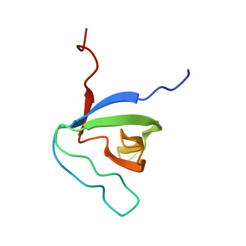
    Transient receptor potential (TRP) channels are polymodally regulated ion channels. TRPV4 (vanilloid 4) is sensitized by PIP 2 and desensitized by Syndapin3/PACSIN3, which bind to the structurally uncharacterized TRPV4 N terminus. We determined the nuclear magnetic resonance structure of the Syndapin3/PACSIN3 SH3 domain in complex with the TRPV4 N-terminal proline-rich region (PRR), which binds as a class I polyproline II (PPII) helix. This PPII conformation is broken by a conserved proline in a cis conformation. Beyond the PPII, we find that the proximal TRPV4 N terminus is unstructured, a feature conserved across species thus explaining the difficulties in resolving it in previous structural studies. Syndapin/PACSIN SH3 domain binding leads to rigidification of both the PRR and the adjacent PIP 2 binding site. We determined the affinities of the TRPV4 N terminus for PACSIN1, 2, and 3 SH3 domains and PIP 2 and deduce a hierarchical interaction network where Syndapin/PACSIN binding influences the PIP 2 binding site but not vice versa.


  • Organizational Affiliation

    Institute for Pharmacy and Biochemistry, Johannes Gutenberg-Universität Mainz, 55128 Mainz, Germany; Center for Biomolecular Magnetic Resonance (BMRZ), Goethe-Universität, 60438 Frankfurt am Main, Germany.


Macromolecules
Find similar proteins by:  (by identity cutoff)  |  3D Structure
Entity ID: 1
MoleculeChains Sequence LengthOrganismDetailsImage
PACSIN 367Gallus gallusMutation(s): 0 
Gene Names: PACSIN3
UniProt
Find proteins for Q1G1I6 (Gallus gallus)
Explore Q1G1I6 
Go to UniProtKB:  Q1G1I6
Entity Groups  
Sequence Clusters30% Identity50% Identity70% Identity90% Identity95% Identity100% Identity
UniProt GroupQ1G1I6
Sequence Annotations
Expand
  • Reference Sequence

Find similar proteins by:  Sequence   |   3D Structure  

Entity ID: 2
MoleculeChains Sequence LengthOrganismDetailsImage
PRR15Homo sapiensMutation(s): 0 
UniProt
Find proteins for A0A1D5PXA5 (Gallus gallus)
Explore A0A1D5PXA5 
Go to UniProtKB:  A0A1D5PXA5
Entity Groups  
Sequence Clusters30% Identity50% Identity70% Identity90% Identity95% Identity100% Identity
UniProt GroupA0A1D5PXA5
Sequence Annotations
Expand
  • Reference Sequence
Experimental Data & Validation

Experimental Data

  • Method: SOLUTION NMR
  • Conformers Calculated: 100 
  • Conformers Submitted: 10 
  • Selection Criteria: structures with the least restraint violations 

Structure Validation

View Full Validation Report


Entry History 

Deposition Data

Revision History  (Full details and data files)

  • Version 1.0: 2018-10-03
    Type: Initial release
  • Version 1.1: 2018-12-19
    Changes: Data collection, Database references
  • Version 1.2: 2019-05-08
    Changes: Data collection