6F36

Polytomella Fo model


Experimental Data Snapshot

  • Method: ELECTRON MICROSCOPY
  • Resolution: 3.70 Å
  • Aggregation State: PARTICLE 
  • Reconstruction Method: SINGLE PARTICLE 

wwPDB Validation   3D Report Full Report


This is version 1.2 of the entry. See complete history


Literature

Structural basis of proton translocation and force generation in mitochondrial ATP synthase.

Klusch, N.Murphy, B.J.Mills, D.J.Yildiz, O.Kuhlbrandt, W.

(2017) Elife 6

  • DOI: https://doi.org/10.7554/eLife.33274
  • Primary Citation of Related Structures:  
    6F36

  • PubMed Abstract: 

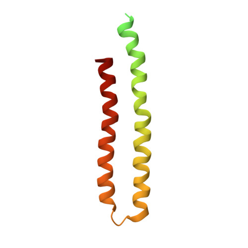
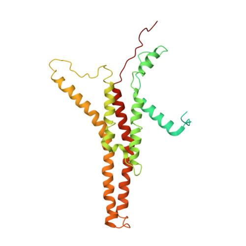
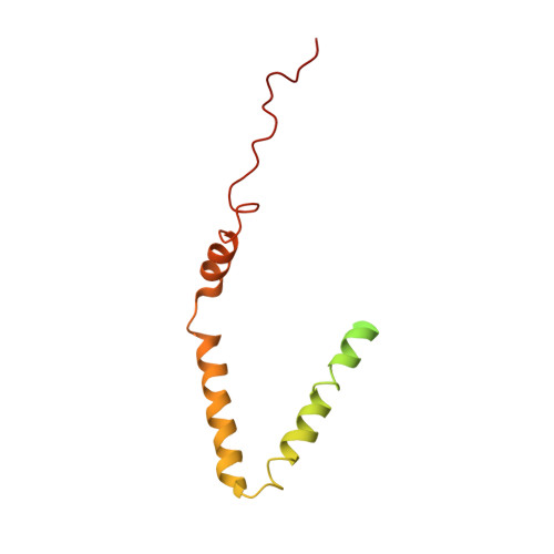
    ATP synthases produce ATP by rotary catalysis, powered by the electrochemical proton gradient across the membrane. Understanding this fundamental process requires an atomic model of the proton pathway. We determined the structure of an intact mitochondrial ATP synthase dimer by electron cryo-microscopy at near-atomic resolution. Charged and polar residues of the a -subunit stator define two aqueous channels, each spanning one half of the membrane. Passing through a conserved membrane-intrinsic helix hairpin, the lumenal channel protonates an acidic glutamate in the c -ring rotor. Upon ring rotation, the protonated glutamate encounters the matrix channel and deprotonates. An arginine between the two channels prevents proton leakage. The steep potential gradient over the sub-nm inter-channel distance exerts a force on the deprotonated glutamate, resulting in net directional rotation.


  • Organizational Affiliation

    Department of Structural Biology, Max Planck Institute of Biophysics, Frankfurt, Germany.


Macromolecules
Find similar proteins by:  (by identity cutoff)  |  3D Structure
Entity ID: 1
MoleculeChains Sequence LengthOrganismDetailsImage
Mitochondrial ATP synthase subunit c
A, B, C, D, E
A, B, C, D, E, F, G, H, I, J
127Polytomella sp. Pringsheim 198.80Mutation(s): 0 
Membrane Entity: Yes 
UniProt
Find proteins for D7P7X5 (Polytomella sp. Pringsheim 198.80)
Explore D7P7X5 
Go to UniProtKB:  D7P7X5
Entity Groups  
Sequence Clusters30% Identity50% Identity70% Identity90% Identity95% Identity100% Identity
UniProt GroupD7P7X5
Sequence Annotations
Expand
  • Reference Sequence
Find similar proteins by:  (by identity cutoff)  |  3D Structure
Entity ID: 2
MoleculeChains Sequence LengthOrganismDetailsImage
Mitochondrial ATP synthase subunit 6K [auth M]327Polytomella sp. Pringsheim 198.80Mutation(s): 0 
Membrane Entity: Yes 
UniProt
Find proteins for H8PGG3 (Polytomella sp. Pringsheim 198.80)
Explore H8PGG3 
Go to UniProtKB:  H8PGG3
Entity Groups  
Sequence Clusters30% Identity50% Identity70% Identity90% Identity95% Identity100% Identity
UniProt GroupH8PGG3
Sequence Annotations
Expand
  • Reference Sequence
Find similar proteins by:  (by identity cutoff)  |  3D Structure
Entity ID: 3
MoleculeChains Sequence LengthOrganismDetailsImage
Mitochondrial ATP synthase subunit ASA6L [auth N]151Polytomella sp. Pringsheim 198.80Mutation(s): 0 
UniProt
Find proteins for D7P897 (Polytomella sp. Pringsheim 198.80)
Explore D7P897 
Go to UniProtKB:  D7P897
Entity Groups  
Sequence Clusters30% Identity50% Identity70% Identity90% Identity95% Identity100% Identity
UniProt GroupD7P897
Sequence Annotations
Expand
  • Reference Sequence
Experimental Data & Validation

Experimental Data

  • Method: ELECTRON MICROSCOPY
  • Resolution: 3.70 Å
  • Aggregation State: PARTICLE 
  • Reconstruction Method: SINGLE PARTICLE 

Structure Validation

View Full Validation Report


Entry History & Funding Information

Deposition Data


Funding OrganizationLocationGrant Number
Max Planck SocietyGermany--
German Research FoundationGermanySFB807
European Molecular Biology OrganizationGermany--

Revision History  (Full details and data files)

  • Version 1.0: 2017-12-20
    Type: Initial release
  • Version 1.1: 2018-01-31
    Changes: Author supporting evidence, Data processing
  • Version 1.2: 2019-12-11
    Changes: Other