6F2C

Methylglyoxal synthase MgsA from Bacillus subtilis


Experimental Data Snapshot

  • Method: X-RAY DIFFRACTION
  • Resolution: 2.34 Å
  • R-Value Free: 0.207 
  • R-Value Work: 0.171 
  • R-Value Observed: 0.172 

wwPDB Validation   3D Report Full Report


This is version 1.4 of the entry. See complete history


Literature

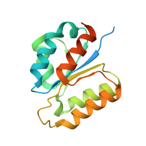
Structural basis for the regulatory interaction of the methylglyoxal synthase MgsA with the carbon flux regulator Crh inBacillus subtilis.

Dickmanns, A.Zschiedrich, C.P.Arens, J.Parfentev, I.Gundlach, J.Hofele, R.Neumann, P.Urlaub, H.Gorke, B.Ficner, R.Stulke, J.

(2018) J Biol Chem 293: 5781-5792

  • DOI: https://doi.org/10.1074/jbc.RA117.001289
  • Primary Citation of Related Structures:  
    6F2C

  • PubMed Abstract: 

    Utilization of energy-rich carbon sources such as glucose is fundamental to the evolutionary success of bacteria. Glucose can be catabolized via glycolysis for feeding the intermediary metabolism. The methylglyoxal synthase MgsA produces methylglyoxal from the glycolytic intermediate dihydroxyacetone phosphate. Methylglyoxal is toxic, requiring stringent regulation of MgsA activity. In the Gram-positive bacterium Bacillus subtilis , an interaction with the phosphoprotein Crh controls MgsA activity. In the absence of preferred carbon sources, Crh is present in the nonphosphorylated state and binds to and thereby inhibits MgsA. To better understand the mechanism of regulation of MgsA, here we performed biochemical and structural analyses of B. subtilis MgsA and of its interaction with Crh. Our results indicated that MgsA forms a hexamer ( i.e. a trimer of dimers) in the crystal structure, whereas it seems to exist in an equilibrium between a dimer and hexamer in solution. In the hexamer, two alternative dimers could be distinguished, but only one appeared to prevail in solution. Further analysis strongly suggested that the hexamer is the biologically active form. In vitro cross-linking studies revealed that Crh interacts with the N-terminal helices of MgsA and that the Crh-MgsA binding inactivates MgsA by distorting and thereby blocking its active site. In summary, our results indicate that dimeric and hexameric MgsA species exist in an equilibrium in solution, that the hexameric species is the active form, and that binding to Crh deforms and blocks the active site in MgsA.


  • Organizational Affiliation

    From the Departments of Molecular Structural Biology and adickma@gwdg.de.


Macromolecules
Find similar proteins by:  (by identity cutoff)  |  3D Structure
Entity ID: 1
MoleculeChains Sequence LengthOrganismDetailsImage
Methylglyoxal synthase160Bacillus subtilis subsp. subtilis str. 168Mutation(s): 0 
Gene Names: mgsAypjFBSU22480
EC: 4.2.3.3
UniProt
Find proteins for P42980 (Bacillus subtilis (strain 168))
Explore P42980 
Go to UniProtKB:  P42980
Entity Groups  
Sequence Clusters30% Identity50% Identity70% Identity90% Identity95% Identity100% Identity
UniProt GroupP42980
Sequence Annotations
Expand
  • Reference Sequence
Small Molecules
Ligands 2 Unique
IDChains Name / Formula / InChI Key2D Diagram3D Interactions
GOL
Query on GOL

Download Ideal Coordinates CCD File 
AA [auth E]
CA [auth H]
M [auth A]
MA [auth L]
N [auth A]
AA [auth E],
CA [auth H],
M [auth A],
MA [auth L],
N [auth A],
S [auth B],
T [auth B],
V [auth C],
Z [auth E]
GLYCEROL
C3 H8 O3
PEDCQBHIVMGVHV-UHFFFAOYSA-N
CL
Query on CL

Download Ideal Coordinates CCD File 
BA [auth H]
DA [auth H]
EA [auth K]
FA [auth G]
GA [auth G]
BA [auth H],
DA [auth H],
EA [auth K],
FA [auth G],
GA [auth G],
HA [auth G],
IA [auth I],
JA [auth I],
KA [auth I],
LA [auth I],
NA [auth L],
O [auth A],
P [auth D],
Q [auth D],
R [auth D],
U [auth F],
W [auth C],
X [auth C],
Y [auth E]
CHLORIDE ION
Cl
VEXZGXHMUGYJMC-UHFFFAOYSA-M
Experimental Data & Validation

Experimental Data

  • Method: X-RAY DIFFRACTION
  • Resolution: 2.34 Å
  • R-Value Free: 0.207 
  • R-Value Work: 0.171 
  • R-Value Observed: 0.172 
  • Space Group: P 21 21 21
Unit Cell:
Length ( Å )Angle ( ˚ )
a = 108.93α = 90
b = 109.71β = 90
c = 199.95γ = 90
Software Package:
Software NamePurpose
PHENIXrefinement
XSCALEdata scaling
PDB_EXTRACTdata extraction
XDSdata reduction
PHASERphasing

Structure Validation

View Full Validation Report


Entry History 

Deposition Data

Revision History  (Full details and data files)

  • Version 1.0: 2018-03-07
    Type: Initial release
  • Version 1.1: 2018-03-21
    Changes: Database references
  • Version 1.2: 2018-03-28
    Changes: Data collection, Database references
  • Version 1.3: 2018-05-02
    Changes: Data collection, Database references
  • Version 1.4: 2024-01-17
    Changes: Data collection, Database references, Refinement description