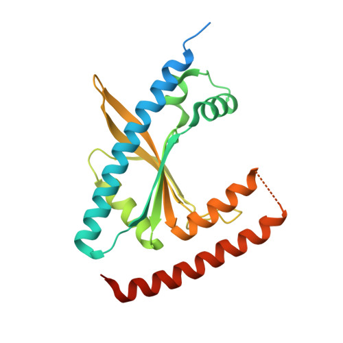
Structural basis for (p)ppGpp synthesis by theStaphylococcus aureussmall alarmone synthetase RelP.
Manav, M.C., Beljantseva, J., Bojer, M.S., Tenson, T., Ingmer, H., Hauryliuk, V., Brodersen, D.E.(2018) J Biol Chem 293: 3254-3264
- PubMed: 29326162
- DOI: https://doi.org/10.1074/jbc.RA117.001374
- Primary Citation of Related Structures:
6EWZ, 6EX0 - PubMed Abstract:
The stringent response is a global reprogramming of bacterial physiology that renders cells more tolerant to antibiotics and induces virulence gene expression in pathogens in response to stress. This process is driven by accumulation of the intracellular alarmone guanosine-5'-di(tri)phosphate-3'-diphosphate ((p)ppGpp), which is produced by enzymes of the RelA SpoT homologue (RSH) family. The Gram-positive Firmicute pathogen, Staphylococcus aureus , encodes three RSH enzymes: a multidomain RSH (Rel) that senses amino acid starvation on the ribosome and two small alarmone synthetase (SAS) enzymes, RelQ (SAS1) and RelP (SAS2). In Bacillus subtilis , RelQ (SAS1) was shown to form a tetramer that is activated by pppGpp and inhibited by single-stranded RNA, but the structural and functional regulation of RelP (SAS2) is unexplored. Here, we present crystal structures of S. aureus RelP in two major functional states, pre-catalytic (bound to GTP and the non-hydrolyzable ATP analogue, adenosine 5'-(α,β-methylene)triphosphate (AMP-CPP)), and post-catalytic (bound to pppGpp). We observed that RelP also forms a tetramer, but unlike RelQ (SAS1), it is strongly inhibited by both pppGpp and ppGpp and is insensitive to inhibition by RNA. We also identified putative metal ion-binding sites at the subunit interfaces that were consistent with the observed activation of the enzyme by Zn 2+ ions. The structures reported here reveal the details of the catalytic mechanism of SAS enzymes and provide a molecular basis for understanding differential regulation of SAS enzymes in Firmicute bacteria.
Organizational Affiliation:
From the Department of Molecular Biology and Genetics, Centre for Bacterial Stress Response and Persistence, Gustav Wieds Vej 10c, DK-8000 Aarhus C, Denmark.


