


Structure of Acinetobacter phage vb_AbaP_AS12 gp42 tailspike
Taylor, N.M.I., Shneider, M.M., Leiman, P.G.To be published.
Experimental Data Snapshot
wwPDB Validation 3D Report Full Report

Entity ID: 1 | |||||
|---|---|---|---|---|---|
| Molecule | Chains | Sequence Length | Organism | Details | Image |
| Tail spike protein | 716 | Acinetobacter phage vB_AbaP_AS12 | Mutation(s): 0 Gene Names: AS12_gp42 | 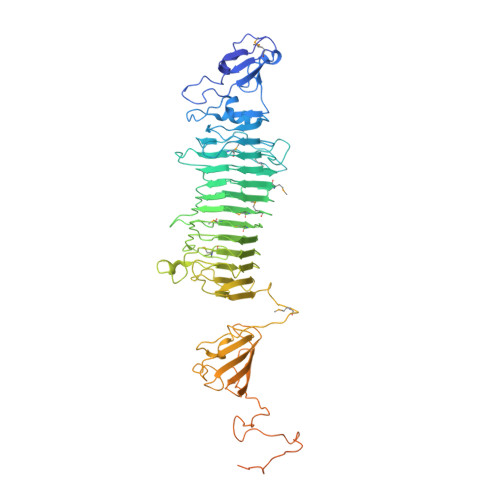 | |
UniProt | |||||
Find proteins for A0A218KRF6 (Acinetobacter phage vB_AbaP_AS12) Explore A0A218KRF6 Go to UniProtKB: A0A218KRF6 | |||||
Entity Groups | |||||
| Sequence Clusters | 30% Identity50% Identity70% Identity90% Identity95% Identity100% Identity | ||||
| UniProt Group | A0A218KRF6 | ||||
Sequence AnnotationsExpand | |||||
| |||||
| Ligands 1 Unique | |||||
|---|---|---|---|---|---|
| ID | Chains | Name / Formula / InChI Key | 2D Diagram | 3D Interactions | |
| ZN Query on ZN | D [auth A] | ZINC ION Zn PTFCDOFLOPIGGS-UHFFFAOYSA-N |  | ||
| Modified Residues 1 Unique | |||||
|---|---|---|---|---|---|
| ID | Chains | Type | Formula | 2D Diagram | Parent |
| MSE Query on MSE | A, B, C | L-PEPTIDE LINKING | C5 H11 N O2 Se |  | MET |
| Length ( Å ) | Angle ( ˚ ) |
|---|---|
| a = 93.202 | α = 90 |
| b = 143.336 | β = 90 |
| c = 176.515 | γ = 90 |
| Software Name | Purpose |
|---|---|
| PHENIX | refinement |
| XDS | data reduction |
| XDS | data scaling |
| SHELXDE | phasing |
| Funding Organization | Location | Grant Number |
|---|---|---|
| EPFL | Switzerland | EPFL 577-1 |
RCSB PDB (citation) is hosted by
RCSB PDB is a member of the

