6ES9

Methylsuccinyl-CoA dehydrogenase of Paracoccus denitrificans with bound flavin adenine dinucleotide


Experimental Data Snapshot

  • Method: X-RAY DIFFRACTION
  • Resolution: 1.37 Å
  • R-Value Free: 0.185 
  • R-Value Work: 0.176 
  • R-Value Observed: 0.176 

wwPDB Validation   3D Report Full Report


Ligand Structure Quality Assessment 


This is version 1.3 of the entry. See complete history


Literature

Structural basis for substrate specificity of methylsuccinyl-CoA dehydrogenase, an unusual member of the acyl-CoA dehydrogenase family.

Schwander, T.McLean, R.Zarzycki, J.Erb, T.J.

(2018) J Biol Chem 293: 1702-1712

  • DOI: https://doi.org/10.1074/jbc.RA117.000764
  • Primary Citation of Related Structures:  
    6ES9

  • PubMed Abstract: 

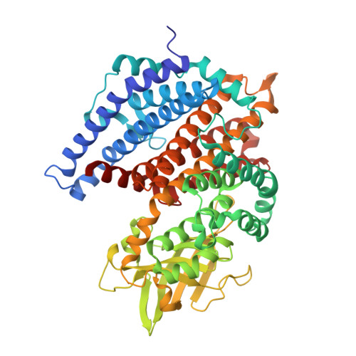
    (2 S )-methylsuccinyl-CoA dehydrogenase (MCD) belongs to the family of FAD-dependent acyl-CoA dehydrogenase (ACD) and is a key enzyme of the ethylmalonyl-CoA pathway for acetate assimilation. It catalyzes the oxidation of (2 S )-methylsuccinyl-CoA to α,β-unsaturated mesaconyl-CoA and shows only about 0.5% activity with succinyl-CoA. Here we report the crystal structure of MCD at a resolution of 1.37 Å. The enzyme forms a homodimer of two 60-kDa subunits. Compared with other ACDs, MCD contains an ∼170-residue-long N-terminal extension that structurally mimics a dimer-dimer interface of these enzymes that are canonically organized as tetramers. MCD catalyzes the unprecedented oxidation of an α-methyl branched dicarboxylic acid CoA thioester. Substrate specificity is achieved by a cluster of three arginines that accommodates the terminal carboxyl group and a dedicated cavity that facilitates binding of the C2 methyl branch. MCD apparently evolved toward preventing the nonspecific oxidation of succinyl-CoA, which is a close structural homolog of (2 S )-methylsuccinyl-CoA and an essential intermediate in central carbon metabolism. For different metabolic engineering and biotechnological applications, however, an enzyme that can oxidize succinyl-CoA to fumaryl-CoA is sought after. Based on the MCD structure, we were able to shift substrate specificity of MCD toward succinyl-CoA through active-site mutagenesis.


  • Organizational Affiliation

    From the Department of Biochemistry and Synthetic Metabolism, Max-Planck-Institute for Terrestrial Microbiology Marburg, Karl-von-Frisch-Strasse 10, D-35043 Marburg, Germany and.


Macromolecules
Find similar proteins by:  (by identity cutoff)  |  3D Structure
Entity ID: 1
MoleculeChains Sequence LengthOrganismDetailsImage
Acyl-CoA dehydrogenase
A, B
551Paracoccus denitrificans PD1222Mutation(s): 0 
Gene Names: Pden_2840
EC: 1.3.8
UniProt
Find proteins for A1B5Y0 (Paracoccus denitrificans (strain Pd 1222))
Explore A1B5Y0 
Go to UniProtKB:  A1B5Y0
Entity Groups  
Sequence Clusters30% Identity50% Identity70% Identity90% Identity95% Identity100% Identity
UniProt GroupA1B5Y0
Sequence Annotations
Expand
  • Reference Sequence
Small Molecules
Ligands 3 Unique
IDChains Name / Formula / InChI Key2D Diagram3D Interactions
FAD
Query on FAD

Download Ideal Coordinates CCD File 
F [auth A],
J [auth B]
FLAVIN-ADENINE DINUCLEOTIDE
C27 H33 N9 O15 P2
VWWQXMAJTJZDQX-UYBVJOGSSA-N
COA
Query on COA

Download Ideal Coordinates CCD File 
E [auth A],
I [auth B]
COENZYME A
C21 H36 N7 O16 P3 S
RGJOEKWQDUBAIZ-IBOSZNHHSA-N
SO4
Query on SO4

Download Ideal Coordinates CCD File 
C [auth A],
D [auth A],
G [auth B],
H [auth B]
SULFATE ION
O4 S
QAOWNCQODCNURD-UHFFFAOYSA-L
Experimental Data & Validation

Experimental Data

  • Method: X-RAY DIFFRACTION
  • Resolution: 1.37 Å
  • R-Value Free: 0.185 
  • R-Value Work: 0.176 
  • R-Value Observed: 0.176 
  • Space Group: P 21 21 21
Unit Cell:
Length ( Å )Angle ( ˚ )
a = 81.596α = 90
b = 105.193β = 90
c = 138.939γ = 90
Software Package:
Software NamePurpose
PHENIXrefinement
SCALAdata scaling
TRUNCATEdata reduction
PHASERphasing
PDB_EXTRACTdata extraction

Structure Validation

View Full Validation Report


Ligand Structure Quality Assessment 


Entry History 

Deposition Data

Revision History  (Full details and data files)

  • Version 1.0: 2018-01-03
    Type: Initial release
  • Version 1.1: 2018-02-14
    Changes: Database references
  • Version 1.2: 2022-07-13
    Changes: Database references, Structure summary
  • Version 1.3: 2024-01-17
    Changes: Data collection, Refinement description